КАТЕГОРИИ:
Архитектура-(3434)Астрономия-(809)Биология-(7483)Биотехнологии-(1457)Военное дело-(14632)Высокие технологии-(1363)География-(913)Геология-(1438)Государство-(451)Демография-(1065)Дом-(47672)Журналистика и СМИ-(912)Изобретательство-(14524)Иностранные языки-(4268)Информатика-(17799)Искусство-(1338)История-(13644)Компьютеры-(11121)Косметика-(55)Кулинария-(373)Культура-(8427)Лингвистика-(374)Литература-(1642)Маркетинг-(23702)Математика-(16968)Машиностроение-(1700)Медицина-(12668)Менеджмент-(24684)Механика-(15423)Науковедение-(506)Образование-(11852)Охрана труда-(3308)Педагогика-(5571)Полиграфия-(1312)Политика-(7869)Право-(5454)Приборостроение-(1369)Программирование-(2801)Производство-(97182)Промышленность-(8706)Психология-(18388)Религия-(3217)Связь-(10668)Сельское хозяйство-(299)Социология-(6455)Спорт-(42831)Строительство-(4793)Торговля-(5050)Транспорт-(2929)Туризм-(1568)Физика-(3942)Философия-(17015)Финансы-(26596)Химия-(22929)Экология-(12095)Экономика-(9961)Электроника-(8441)Электротехника-(4623)Энергетика-(12629)Юриспруденция-(1492)Ядерная техника-(1748)
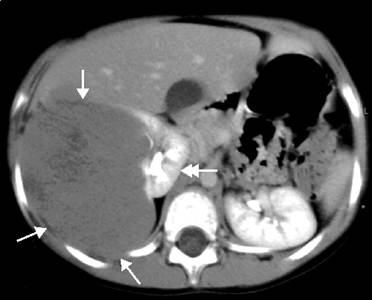
Diagnosis. After an abdominal mass is identified, radiographic imaging is performed to determine the anatomic location and extent of the mass
|
|
|
|
After an abdominal mass is identified, radiographic imaging is performed to determine the anatomic location and extent of the mass.
The diagnostic workup includes a chest x-ray and an abdominal ultrasound. The chest x-ray is done to evaluate for the presence of pulmonary metastasis. Additional studies include urinalysis, abdominal plain film, and computed tomography (Image 9.18). The urine may contain red blood cells (20% of cases) or hyaluronic acid. The abdominal x-ray may show “eggshell” calcifications. This is in contrast to the “speckled” calcifications seen in neuroblastoma and the “popcorn” calcifications seen in teratomas.
|
| Image 9.18 CT scan in a patient with a right-sided Wilms` tumor with favorable histology. |
Although magnetic resonance imaging (MRI) avoids radiation exposure, it has not been shown to be superior to CT scanning in standard assessments. MRI is currently being evaluated as a method to help distinguish nephrogenic rests from WT and may be the preferred method to follow children with bilateral WT after resection.
F-fluorodeoxyglucose positron emission tomography (FDG PET) has not been fully delineated in pediatric cancers. It is recognized that FDG PET has an established role in Hodgkin lymphoma and increasingly in sarcomas in children, but its role in WT is unclear.
|
|
|
|
|
Дата добавления: 2014-10-15; Просмотров: 259; Нарушение авторских прав?; Мы поможем в написании вашей работы!