КАТЕГОРИИ:
Архитектура-(3434)Астрономия-(809)Биология-(7483)Биотехнологии-(1457)Военное дело-(14632)Высокие технологии-(1363)География-(913)Геология-(1438)Государство-(451)Демография-(1065)Дом-(47672)Журналистика и СМИ-(912)Изобретательство-(14524)Иностранные языки-(4268)Информатика-(17799)Искусство-(1338)История-(13644)Компьютеры-(11121)Косметика-(55)Кулинария-(373)Культура-(8427)Лингвистика-(374)Литература-(1642)Маркетинг-(23702)Математика-(16968)Машиностроение-(1700)Медицина-(12668)Менеджмент-(24684)Механика-(15423)Науковедение-(506)Образование-(11852)Охрана труда-(3308)Педагогика-(5571)Полиграфия-(1312)Политика-(7869)Право-(5454)Приборостроение-(1369)Программирование-(2801)Производство-(97182)Промышленность-(8706)Психология-(18388)Религия-(3217)Связь-(10668)Сельское хозяйство-(299)Социология-(6455)Спорт-(42831)Строительство-(4793)Торговля-(5050)Транспорт-(2929)Туризм-(1568)Физика-(3942)Философия-(17015)Финансы-(26596)Химия-(22929)Экология-(12095)Экономика-(9961)Электроника-(8441)Электротехника-(4623)Энергетика-(12629)Юриспруденция-(1492)Ядерная техника-(1748)
Sacrococcygeal Teratoma
|
|
|
|
Etiology
Incidence
Localization
The kind of pluripotent cell appears to be unimportant, apart from constraining the location of the teratoma in the body.
Whereas in adults 90% of germ cell tumors are at gonadal locations, in childhood, the extragonadal site is more common until puberty, at which time the gonadal sites are more common.
Teratomas derived from embryonal cells usually occur on the body midline: in the brain, elsewhere inside the skull, in the nose, in the tongue, under the tongue, and in the neck (cervical teratoma), mediastinum, retroperitoneum, and attached to the coccyx. However, teratomas may also occur elsewhere: very rarely in solid organs (most notably the heart and liver) and hollow organs (such as the stomach and bladder), and more commonly on the skull sutures.
Embryonal teratomas most commonly occur in the sacrococcygeal region: sacrococcygeal teratoma is the single most common tumor found in newborn babies.
Of teratomas on the skull sutures, approximately 50% are found in or adjacent to the orbit.
Teratomas are interesting but uncommon lesions, probably occurring in 1 in 20,000-40,000 live births. The exact incidence is difficult to ascertain.
The anatomic distribution of these lesions varies between reporting institutions, but the sacrococcygeal lesion appears to be most common.
Sacrococcygeal teratomas account for the majority of cases (45-65%). The next most common locations are: gonadal (10-35%), mediastinal (10-12%), retroperitoneal (3-5%), cervical (3-6%), presacral (3-5%), and central nervous system (2-4%).
Approximately 20% of pediatric germ cell tumors are malignant, and they represent 1% to 3% of all malignant tumors in childhood and adolescence.
Teratomas arise from germ cells or other totipotential cells. Primordial germ cells appear during the third week of gestation in the wall of the yolk sac near the allantois. They move along the dorsal mesentery of the hindgut, reaching the genital ridges by about the sixth week of gestation. Germ cells that do not complete this journey can develop into teratomas. While the totipotential nature of germ cells and their path of migration explain the location and pathology of the more common teratomas (sacroccygeal and gonadal), intracranial and mediastinal locations are more difficult to explain.
Tumors of the sacrococcygeal region, referred to as sacrococcygeal teratomas (SCTs) in most reports, generally present in two distinct fashions: neonates with large predominantly external lesions, which are detected in utero or at birth and are rarely malignant; and older infants and children who present with primarily hidden pelvic tumors with a much higher rate of malignancy. SCTs are the most common extragonadal tumor in neonates, accounting for up to 70% of all teratomas in childhood. Interestingly, 75% occur in females.
On examination, the visible portion of the lesion is skin covered and posterior to the anus (Image 9.19). In some patients all or part of the lesion may be in the retrorectal space and/or the retroperitoneum. In these cases, patients will present with rectal pain, constipation, and/or a mass.
Associated anomalies occur in 10-15% of cases and include imperforate anus, anorectal stenosis, anorectal agenesis, sacral hemivertebra, absence of the sacrum and coccyx, and anterior meningocele. Currarinos triad is the association of a pre-sacral mass with anorectal stenosis and a sacral deformity.
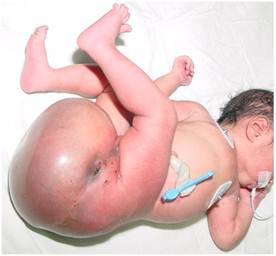
| Image 9.19 Sacrococcygeal teratoma. |
|
|
|
|
|
Дата добавления: 2014-10-15; Просмотров: 271; Нарушение авторских прав?; Мы поможем в написании вашей работы!