КАТЕГОРИИ:
Архитектура-(3434)Астрономия-(809)Биология-(7483)Биотехнологии-(1457)Военное дело-(14632)Высокие технологии-(1363)География-(913)Геология-(1438)Государство-(451)Демография-(1065)Дом-(47672)Журналистика и СМИ-(912)Изобретательство-(14524)Иностранные языки-(4268)Информатика-(17799)Искусство-(1338)История-(13644)Компьютеры-(11121)Косметика-(55)Кулинария-(373)Культура-(8427)Лингвистика-(374)Литература-(1642)Маркетинг-(23702)Математика-(16968)Машиностроение-(1700)Медицина-(12668)Менеджмент-(24684)Механика-(15423)Науковедение-(506)Образование-(11852)Охрана труда-(3308)Педагогика-(5571)Полиграфия-(1312)Политика-(7869)Право-(5454)Приборостроение-(1369)Программирование-(2801)Производство-(97182)Промышленность-(8706)Психология-(18388)Религия-(3217)Связь-(10668)Сельское хозяйство-(299)Социология-(6455)Спорт-(42831)Строительство-(4793)Торговля-(5050)Транспорт-(2929)Туризм-(1568)Физика-(3942)Философия-(17015)Финансы-(26596)Химия-(22929)Экология-(12095)Экономика-(9961)Электроника-(8441)Электротехника-(4623)Энергетика-(12629)Юриспруденция-(1492)Ядерная техника-(1748)
Staging
|
|
|
|
Classification
Differential Diagnosis
Diagnosis
At present there is no specific laboratory test for HL. An excisional biopsy of a suspicious lymph node should be the initial step to diagnosis.
Diagnosis depends on histologic evaluation and identification of Reed-Sternberg cells, hence biopsy is mandatory. An adequate amount of tissue should be excised for analysis and, if possible, several lymph nodes should be evaluated histologically. In the absence of palpable peripheral lymph nodes in a child with mediastinal disease, mediastinoscopy or thoracoscopy/thoracotomy are necessary to obtain tissue for diagnosis.
The differential diagnosis includes systemic infections, infectious lymphadenopathy, NHL, Langerhan's histiocytosis, mononucleosis and other causes of generalized or localized lymphadenopathy.
Any child with unexplained lymphadenopathy should receive a careful history and physical examination and a chest radiograph to rule out the presence of a mediastinal mass prior to lymph node biopsy.
The current World Health Organization classification system separates HL into two broad categories:
v Classical
v Lymphocyte predominant
Classical HL has four subtypes:
· lymphocyte depleted,
· nodular sclerosing,
· mixed cellularity, and
· classical lymphocyte rich.
Classical HL accounts for 90% of all cases. For children, nodular sclerosis is the most common subtype, accounting for 65% of cases.
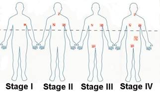
Staging has both clinical and pathologic features. The Ann Arbor staging system and its Cotswolds modification remain the standard for adult and pediatric HL (Table 9.5).
Clinical staging is dependent on: (1) involvement of single or multiple lymph node regions, (2) involvement of single or multiple extra-lymphatic organs, and (3) presence of disease on one or both sides of the diaphragm.
Table 9.5
Hodgkin Lymphoma Staging:
Ann Arbor Classification with Cotswolds Modification
| Stage I - Involvement of a single lymph node region or lymphoid structure (e.g., spleen, thymus, Waldeyer ring) or involvement of a single extralymphatic site |
| Stage II - Involvement of two or more lymph node regions on the same side of the diaphragm |
| Stage III - Indicates that the cancer has spread to both sides of the diaphragm, including one organ or area near the lymph nodes or the spleen With or without involvement of splenic, hilar, celiac, or portal nodes; with involvement of paraaortic, iliac, and mesenteric nodes |
| Stage IV - Indicates that the cancer has spread to both sides of the diaphragm, including one organ or area near the lymph nodes or the spleen |
|
Modifiers:
A or B: The absence of constitutional (B-type) symptoms is denoted by adding an "A" to the stage; the presence is denoted by adding a "B" to the stage.
E: Used if the disease is "extranodal" (not in the lymph nodes) or has spread from lymph nodes to adjacent tissue.
X: Used if the largest deposit is greater than 10 cm large (bulky disease), or whether the mediastinum is wider than one third of the chest on a chest x-ray.
S: Used if the disease has spread to the spleen.
The nature of the staging is (occasionally) expressed with:
CS: Clinical stage as obtained by doctor's examinations and tests.
PS: Pathologic stage as obtained by exploratory laparotomy (surgery performed through an abdominal incision) with splenectomy (surgical removalof the spleen). Note: Exploratory laparotomy has fallen out of favor for lymphoma staging.
|
|
|
|
|
Дата добавления: 2014-10-15; Просмотров: 313; Нарушение авторских прав?; Мы поможем в написании вашей работы!