КАТЕГОРИИ:
Архитектура-(3434)Астрономия-(809)Биология-(7483)Биотехнологии-(1457)Военное дело-(14632)Высокие технологии-(1363)География-(913)Геология-(1438)Государство-(451)Демография-(1065)Дом-(47672)Журналистика и СМИ-(912)Изобретательство-(14524)Иностранные языки-(4268)Информатика-(17799)Искусство-(1338)История-(13644)Компьютеры-(11121)Косметика-(55)Кулинария-(373)Культура-(8427)Лингвистика-(374)Литература-(1642)Маркетинг-(23702)Математика-(16968)Машиностроение-(1700)Медицина-(12668)Менеджмент-(24684)Механика-(15423)Науковедение-(506)Образование-(11852)Охрана труда-(3308)Педагогика-(5571)Полиграфия-(1312)Политика-(7869)Право-(5454)Приборостроение-(1369)Программирование-(2801)Производство-(97182)Промышленность-(8706)Психология-(18388)Религия-(3217)Связь-(10668)Сельское хозяйство-(299)Социология-(6455)Спорт-(42831)Строительство-(4793)Торговля-(5050)Транспорт-(2929)Туризм-(1568)Физика-(3942)Философия-(17015)Финансы-(26596)Химия-(22929)Экология-(12095)Экономика-(9961)Электроника-(8441)Электротехника-(4623)Энергетика-(12629)Юриспруденция-(1492)Ядерная техника-(1748)
Исходят при неожиданном растяжении или вращательных ваются при чрезмерных нагрузках и угловых деформациях 2 страница
1 - Palmar ligaments; 2 - Distal phalanx; 3 - Proximal phalanx; 4 - Metacarpals [I-V]; 5 - Palmar carpometacarpal ligaments; 6 - Tubercle; 7 - Radial collateral ligament of wrist joint; 8 - Radial styloid process; 9 - Palmar radiocarpal ligament; 10 - Palmar radioulnar ligament; 11 - Radius; 12 - Ulna; 13 - Distal radio-ulnar joint; 14 - Ulnar styloid process; 15 - Palmar ulnocarpal ligament; 16 - Flexor carpi ulnaris; Tendon; 17 - Pisiform; 18 - Palmar intercarpal ligaments; 19 - Hook of hamate; 20 - Palmar metacarpal ligaments; 21 - Metacarpophalangeal joints, collateral ligaments; 22 - Deep transverse metacarpal ligament; 23 - Middle phalanx; 24 - Proximal interphalangeal joint; Joint capsule; Articular capsule; 25 - Distal interphalangeae joint; Joint capsule; Articular capsule
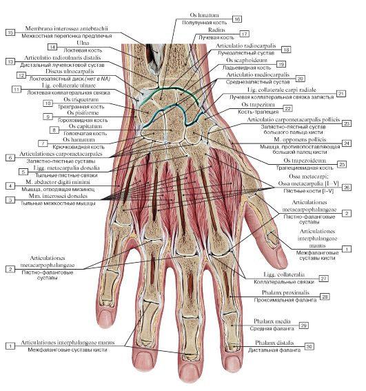 Рис. 194. Фронтальный распил кисти, правой:
1 - Interphalangeal joints of hand; 2 - Metacarpophalangeal joints; 3 - Dorsal interossei; 4 - Abductor digiti minimi; 5 - Dorsal metacarpal ligaments; 6 - Carpometacarpal joints; 7 - Hamate; 8 - Capitate; 9 - Pisiform; 10 - Triquetrum; 11 - Ulnar collateral ligament; 12 - Ulnocarpal disc; 13 - Distal radio-ulnar joint; 14 - Ulna; 15 - Interosseous membrane of forearm; 16 - Lunate; 17 - Radius; 18 - Wrist joint; 19 - Scaphoid; 20 - Midcarpal joint; 21 - Radial collateral ligament of wrist jont; 22 - Trapezium; 23 - Carpometacarpal joint of thumb; 24 - Opponens pollicis; 25 - Trapezoid; 26 - Metacarpals [I-V]; 27 - Collateral ligaments; 28 - Proximal phalanx;
29 - Middle phalanx; 30 - Distal phalanx
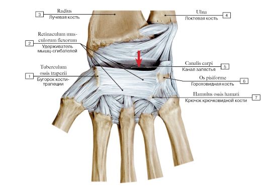 Рис. 195. Удерживатель сгибателей и канал запястья правой кисти, общий вид, вид спереди:
1 - Tubercle; 2 - Flexor retinaculum; 3 - Radius; 4 - Ulna; 5 - Carpal tunnel; 6 - Pisiform; 7 - Hook of hamate
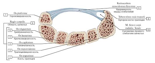 Рис. 196. Канал запястья:
1 - Trapezium; 2 - Trapezoid; 3 - Capitate; 4 - Hamate; 5 - Triquetrum; 6 = 1 + 2 + 3 + 4 + 5 - Carpal region; 6 - Pisiform; 8 - Flexor retinaculum; 9 - Tubercle; 10 - Flexor carpi radialis; Tendon
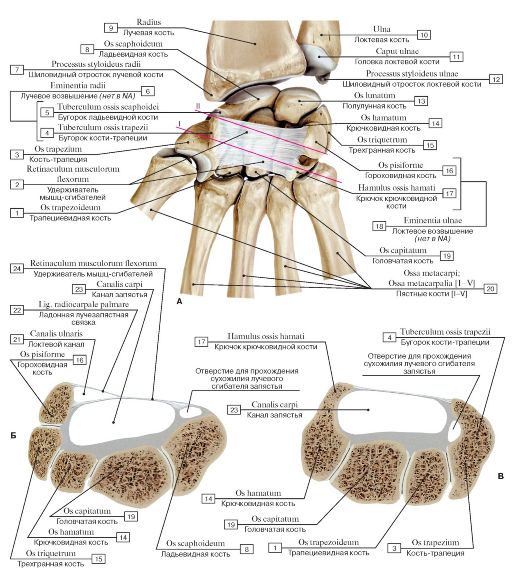 Рис. 197. Удерживатель сгибателей и канал запястья кисти, правой (А - вид спереди, все остальные ткани удалены; Б - поперечный распил кисти на уровне проксимального ряда костей запястья - линия I; В - поперечный распил кисти на уровне дистального ряда костей запястья - линия II):
1 - Trapezoid; 2 - Flexor retinaculum; 3 - Trapezium; 4 - Tubercle of trapezium; 5 - Tubercle of scaphoid; 6 - Radial eminence; 7 - Radial styloid process; 8 - Scaphoid; 9 - Radius; 10 - Ulna; 11 - Head of ulna; 12 - Ulnar styloid process; 13 - Lunate; 14 - Hamate; 15 - Triquetrum; 16 - Pisiform; 17 - Hook of hamate; 18 - Ulnar eminence; 19 - Capitate; 20 - Metacarpals [I-V]; 21 - Ulnar canal; 22 - Palmar radiocarpal ligament; 23 - Carpal tunnel; 24 - Flexor retinaculum
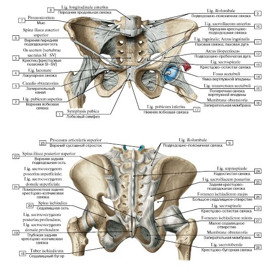 Рис. 198. Соединения костей пояса нижних конечностей и тазобедренные суставы (А - вид спереди, Б - вид сзади):
1 - Pubic symphysis; 2 - Superior pubic ligament; 3 - Obturator canal; 4 - Lacunar ligament; 5 - Sacrum [sacral vertebrae SI-SV]; 6 - Anterior superior iliac spine; 7 - Promontory; 8 - Anterior longitudinal ligament; 9 - Iliolumbar ligament; 10 - Anterior sacro-iliac ligament; 11 - Inguinal ligament; 12 - Iliopectineal arch; 13 - Sacrospinous ligament; 14 - Acetabular fossa; 15 - Transverse acetabular ligament; 16 - Obturator membrane; 17 - Inferior pubic ligament; 18 - Ischial tuberosity; 19 - Deep posterior sacrococcygeal ligament; 20 - Ischial spine; 21 - Superficial posterior sacrococcygeal ligament; 22 - Posterior superior iliac spine; 23 - Superior articular process; 24 - Supraspinous ligament; 25 - Posterior sacro-iliac ligament; 26 - Greater sciatic foramen; 27 - Lesser sciatic foramen; 28 - Sacrotube-
rous ligament
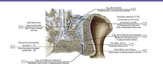 Рис. 199. Крестцово-подвздошное соединение:
1 - Anterior sacro-iliac ligament; 2 - Sacrum [sacral vertebrae I-V]; 3 - Zygapophysial joint; 4 - Iliolumbar ligament; 5 - Lumbar vertebra [LV], costal process; 6 - Interosseous sacro-iliac ligament; 7 - Ilium; 8 - Sacro-iliac joint
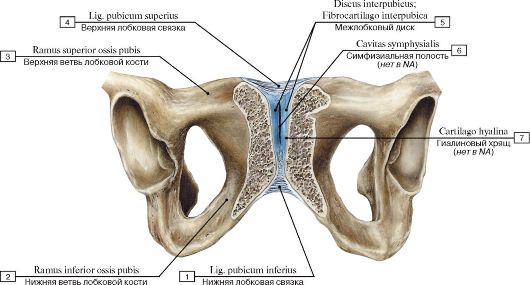 Рис. 200. Лобковый симфиз, вид спереди:
1 - Inferior pubic ligament; 2 - Inferior pubic ramus; 3 - Superior pubic ramus; 4 - Superior pubic ligament; 5 - Interpubic disc; Interpubic fibrocartilage; 6 - Symphysial cavity; 7 - Hyaline cartilage
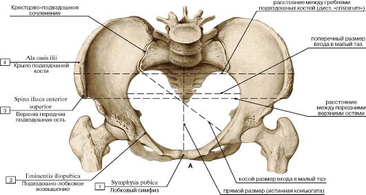
 Рис. 201. Таз женский. Размеры большого и малого таза (А - вид сверху, В - сагиттальный распил, вид изнутри,
со стороны полости таза):
1 - Pubic symphysis; 2 - Iliopubic eminence; 3 - Anterior superior iliac spine; 4 - Ala of ilium; Wing of ilium; 5 - External conjugate; 6 - True conjugate; 7 - External conjugate; 8 - Ischial spine
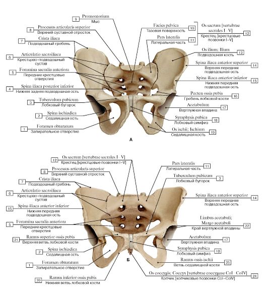 Рис. 202. Таз, вид спереди (А - мужской, Б - женский):
1 - Obturator foramen; 2 - Ischial spine; 3 - Pubic tubercle; 4 - Posterior inferior iliac spine; 5 - Anterior sacral foramina; 6 - Sacro-iliac joint; 7 - Iliac crest; 8 - Superior articular process; 9 - Promontory; 10 - Pelvic surface; 11 - Lateral part; 12 = 10 + 11 - Sacrum [sacral vertebrae I-V];13 - Ilium; 14 - Anterior superior iliac spine; 15 - Anterior inferior iliac spine; 16 - Pecten pubis; Pectineal line; 17 - Acetabulum; 18 - Pubic symphysis; 19 - Ischium; 20 - Inferior pubic ramus; 21 - Superior pubic ramus; 22 - Acetabular margin; 23 - Ramus; 24 - Coccyx
[coccygeal vertebrae CoI-CoIV]
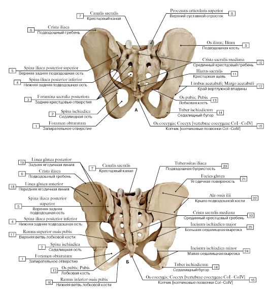 Рис. 203. Таз, вид сзади (А - мужской, Б - женский):
1 - Obturator foramen; 2 - Ischial spine; 3 - Posterior sacral foramina; 4 - Posterior inferior iliac spine; 5 - Posterior superior iliac spine; 6 - Iliac crest; 7 - Sacral canal; 8 - Superior articular process; 9 - Ilium; 10 - Median sacral crest; 11 - Sacral hiatus; 12 - Acetabular margin; 13 - Pubis; 14 - Ischial tuberosity; 15 - Coccyx [coccygeal vertebrae CoI-CoIV]; 16 - Inferior pubic ramus; 17 - Superior pubic ramus; 18 - Anterior gluteal line; 19 - Posterior gluteal line; 20 - Iliac tuberosity; 21 - Gluteal surface; 22 - Ala of ilium; Wing of ilium;
23 - Greater sciatic notch; 24 - Lesser sciatic notch
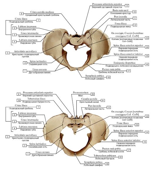 Рис. 204. Таз, вид сверху (А - мужской, Б - женский):
1 - Arcuate line; 2 - Ischial spine; 3 - Sacro-iliac joint; 4 - Outer lip; 5 - Intermediate zone; 6 - Inner lip; 7 = 4 + 5 + 6 - Iliac crest; 8 - Median sacral crest; 9 - Superior articular process; 10 - Base of sacrum; 11 - Lateral part; 12 - Iliac fossa; 13 - Coccyx [coccygeal vertebrae CoI-CoIV]; 14 - Anterior superior iliac spine; 15 - Anterior inferior iliac spine; 16 - Iliopubic eminence; 17 - Pecten pubis; Pectineal line; 18 - Pubic symphysis; 19 - Iliac tuberosity; 20 - Promontory; 21 - Sacral canal; 22 - Pubic tubercle
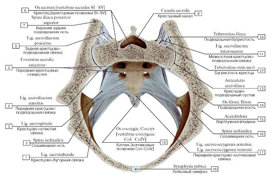 Рис. 205. Связки крестцово-подвздошного сустава, вид сверху, косой распил:
1 - Sacrotuberous ligament; 2 - Ischial spine; 3 - Sacrospinous ligament; 4 - Anterior sacro-iliac ligament; 5 - Anterior sacral foramina; 6 - Posterior sacro-iliac ligament; 7 - Posterior superior iliac spine; 8 - Sacrum [sacral vertebrae SI-SV]; 9 - Sacral canal; 10 - Iliac tuberosity; 11 - Interosseous sacro-iliac ligament; 12 - Sacral tuberosity; 13 - Sacro-iliac joint; 14 - Ilium; 15 - Acetabulum; 16 - Ischial spine; 17 - Anterior sacrococcygeal ligament; 18 - Pubic symphysis; 19 - Coccyx [coccygeal vertebrae CoI-CoIV]
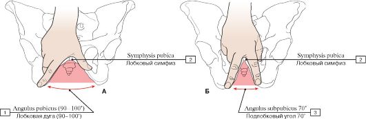 Рис. 206. Лобковая дуга женщины (А), подлобковый угол мужчины (Б):
1 - Pubic arch (90-100°); 2 - Pubic symphysis; 3 - Subpubic angle 70°
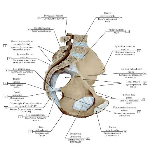 Рис. 207. Связки таза, правая половина таза, вид изнутри, справа:
I - Ischial tuberosity; 2 - Sacrotuberous ligament; 3 - Coccyx [coccygeal vertebrae CoI-CoIV]; 4 - Ischial spine; 5 - Sacral hiatus; 6 - Sacrospinous ligament; 7 - Anterior sacro-iliac ligament; 8 - Sacrum [sacral vertebrae SI-SV]; 9 - Sacral canal; 10 - Spinous process;
II - Intervertebral disc; 12 - Promontory; 13 - Anterior superior iliac spine; 14 - Greater sciatic foramen; 15 - Arcuate line; 16 - Pecten
pubis; Pectineal line; 17 - Lesser sciatic foramen; 18 - Symphysial surface; 19 - Obturator membrane
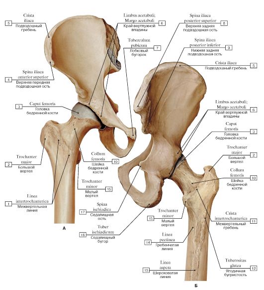 Рис. 208. Взаимоотношения тазовой и бедренной костей, правых (А - вид спереди, Б - вид сзади):
1 - Intertrochanteric line; 2 - Greater trochanter; 3 - Head of femur; 4 - Anterior superior iliac spine; 5 - Iliac crest; 6 - Acetabular margin; 7 - Pubic tubercle; 8 - Posterior superior iliac spine; 9 - Posterior inferior iliac spine; 10 - Neck of femur; 11 - Intertrochanteric crest; 12 - Gluteal tuberosity; 13 - Linea aspera; 14 - Pectineal line; Spiral line; 15- Lesser trochanter; 16 - Ischial tuberosity; 17 - Ischial
spine
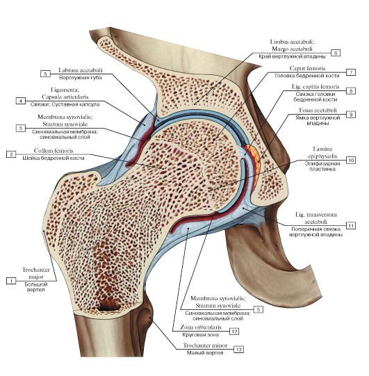 Рис. 209. Тазобедренный сустав, правый, распил во фронтальной плоскости:
1 - Greater trochanter; 2 - Neck of femur; 3 - Synovial membrane; Synovial layer; 4 - Ligaments; Joint capsule; Articular capsule; 5 - Acetabular labrum; 6 - Acetabular margin; 7 - Head of femur; 8 - Ligament of head of femur; 9 - Acetabular fossa; 10 - Epiphysial plate; Growth plate; 11 - Transverse acetabular ligament; 12 - Zona orbicularis; 13 - Lesser trochanter
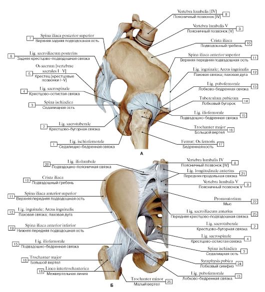 Рис. 210. Связки тазобедренного сустава, правого (А - вид сбоку, справа, Б - вид спереди):
I - Ischiofemoral ligament; 2 - Sacrotuberous ligament; 3 - Ischial spine; 4 - Sacrospinous ligament; 5 - Sacrum [sacral vertebrae I-V]; 6 - Posterior sacro-iliac ligament; 7 - Posterior superior iliac spine; 8 - Lumbar vertebra [IV]; 9 - Lumbar vertebra [V]; 10 - Iliac crest;
II - Anterior superior iliac spine; 12 - Inguinal ligament; 13 - Pubofemoral ligament; 14 - Pubic tubercle; 15 - lliofemoral ligament; 16 - Greater trochanter; 17 - Femur; Thigh bone; 18 - Intertrochanteric line; 19 - Anterior inferior iliac spine; 20 - Iliolumbar ligament;
21 - Anterior longitudinal ligament; 22 - Promontory; 23 - Anterior sacro-iliac ligament; 24 - Pubic symphysis; 25 - Lesser trochanter
 Рис. 211. Связки тазобедренного сустава, правого, вид сзади:
1 - Ischial tuberosity; 2 - Sacrotuberous ligament; 3 - Sacrospinous ligament; 4 - Ischial spine; 5 - Posterior sacro-iliac ligament; 6 - Lumbar vertebrae [V]; 7 - Lumbar vertebrae [IV]; 8 - Iliolumbar ligament; 9 - Iliac crest; 10 - Posterior superior iliac spine; 11 - Iliofemoral ligament; 12 - Greater trochanter; 13 - Intertrochanteric crest; 14 - Lesser trochanter; 15 - Ischiofemoral ligament
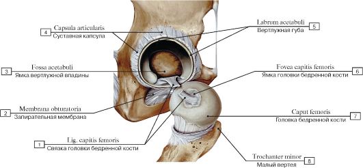 Рис. 212. Связка головки бедренной кости, правой:
1 - Ligament of head of femur; 2 - Obturator membrane; 3 - Acetabular fossa; 4 - Joint capsule; Articular capsule; 5 - Acetabular labrum; 6 - Fovea for ligament of head; 7 - Head of femur; 8 - Lesser trochanter
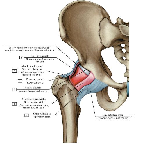 Рис. 213. Места прикрепления суставной капсулы тазобедренного сустава, правого:
1 - Zona orbicularis; 2 - Synovial membrane; Synovial layer; 3 - Head of femur; 4 - Fibrous layer; Fibrous membrane; 5 - lliofemoral ligament; 6 - Pubofemoral ligament
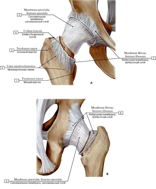 Рис. 214. Связки тазобедренного сустава, правого (А - суставная капсула вскрыта, вид спереди, Б - суставная капсула вскрыта, вид сзади):
1 - Lesser trochanter; 2 - Intertrochanteric line; 3 - Iliac crest; 4 - Neck of femur; 5 - Synovial membrane; Synovial layer; 6 - Fibrous
layer; Fibrous membrane
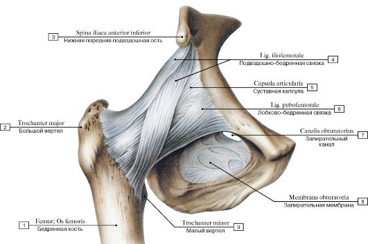 Рис. 215. Связки тазобедренного сустава, правого, вид спереди:
1 - Femur; Thigh bone; 2 - Greater trochanter; 3 - Anterior inferior iliac spine; 4 - Iliofemoral ligament; 5 - Joint capsule; Articular capsule; 6 - Pubofemoral ligament; 7 - Obturator canal; 8 - Obturator membrane; 9 - Lesser trochanter
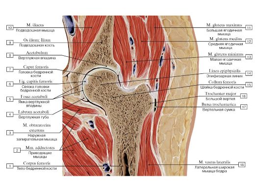 Рис. 216. Топографические взаимоотношения структур таза и верхней трети бедра, правого. Фронтальный распил через тазобедренный сустав:
1 - Shaft of femur; Body of femur; 2 - Adductores; 3 - Obturator externus; 4 - Acetabular labrum; 5 - Acetabular fossa; 6 - Ligament of head of femur; 7 - Head of femur; 8 - Acetabulum; 9 - Ilium; 10 - Iliacus; 11 - Gluteus maximus; 12 - Gluteus medius; 13 - Gluteus minimus; 14 - Epiphysial line; 15 - Neck of femur; 16 - Greater trochanter; 17 - Trochanteric bursa; 18 -Vastus lateralis
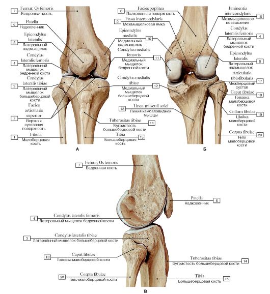 Рис. 217. Кости бедра и голени, формирующие коленный сустав, правый (А - вид спереди, Б - вид сзади, В - вид сбоку, с латеральной стороны):
1 - Fibula; 2 - Superior articular facet; 3 - Lateral condyle of tibia; 4 - Lateral condyle of femur; 5 - Lateral epicondyle; 6 - Patella; 7 - Femur; Thigh bone; 8 - Popliteal surface; 9 - Intercondylar fossa; 10 - Medial epicondyle; 11 - Medial condyle of femur; 12 - Medial condyle of tibia; 13 - Soleal line; 14 - Tibial tuberosity; 15 - Tibia; 16 - Intercondylar eminence; 17 - Tibiofibular joint; Superior tibiofibular joint; 18 - Head of fibula; 19 - Neck of fibula; 20 - Shaft of fibula; Body of fibula
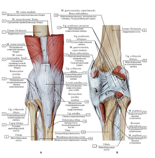 Рис. 218. Коленный сустав, правый (А - вид спереди, Б - вид сзади):
I - Fibula; 2 - Head of fibula; 3 - Tibial collateral ligament; 4 - Lateral patellar retinaculum; 5 -Vastus lateralis; Tendon; 6 -Vastus lateralis; 7 - Femur; Thigh bone; 8 - Rectus femoris; Tendon; 9 -Vastus medialis; 10 - Gastrocnemius, lateral head; Subtendinous bursa;
II - Arcuate popliteal ligament; 12 - Oblique popliteal ligament; 13 - Gastrocnemius, medial head; Subtendinous bursa; 14 - Tibial collateral ligament; 15 - Location of patella; 16 - Medial patellar retinaculum; 17 - Patellar ligament; 18 - Tibial tuberosity; 19 - Tibia;
20 - Interosseous membrane of leg; 21 - Fibular collateral ligament; 22 - Popliteus; 23 - Subpopliteal recess
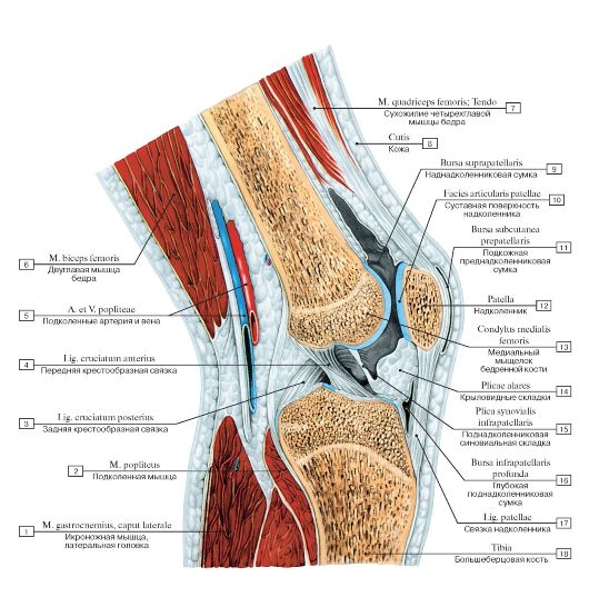 Рис. 219. Коленный сустав, правый, сагиттальный распил:
1 - Gastrocnemius, lateral head; 2 - Popliteus; 3 - Posterior cruciate ligament; 4 - Anterior cruciate ligament; 5 - Popliteal artery and vein; 6 - Biceps femoris; 7 - Quadriceps femoris; Tendon; 8 - Skin; 9 - Suprapatellar bursa; 10 - Articular surface of patella; 11 - Subcutaneous prepatellar bursa; 12 - Patella; 13 - Medial condyle of femur; 14 - Alar folds; 15 - Infrapatellar synovial fold; 16 - Deep infrapatellar bursa;
17 - Patellar ligament; 18 - Tibia
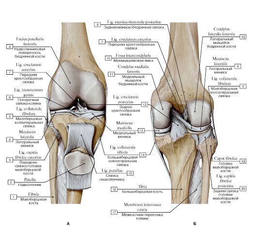 Рис. 220. Внутрисуставные связки коленного сустава, правого (А -вид спереди, надколенник оттянут вниз, Б - вид сзади):
I - Fibula; 2 - Patella; 3 - Anterior ligament of fibular head; 4 - Lateral meniscus; 5 - Fibular collateral ligament; 6 - Transverse ligament of knee; 7 - Anterior cruciate ligament; 8 - Facies patellaris femur; 9 - Posterior meniscofemoral ligament; 10 - Intercondylar fossa;
II - Medial condyle of femur; 12 - Posterior cruciate ligament; 13 - Medial meniscus; 14 - Tibial collateral ligament; 15 - Patellar ligament; 16 - Tibia; 17 - Interosseous membrane of leg; 18 - Lateral condyle of femur; 19 - Head of fibula; 20 - Posterior ligament of fibular
head
 Рис. 221. Связки коленного сустава, правого (А - вид с медиальной стороны, Б - вид с латеральной стороны):
1 - Medial surface of tibia; 2 - Tibial collateral ligament; 3 - Medial meniscus; 4 - Patellar ligament; 5 - Quadratus femoris; 6 - Femur; Thigh bone; 7 - Medial condyle of femur; 8 - Medial epicondyle; 9 - Quadriceps femoris; Tendon; 10 - Lateral epicondyle; 11 - Lateral condyle of femur; 12 - Patella; 13 - Patellar surface; 14 - Lateral meniscus; 15 - Fibular collateral ligament; 16 - Anterior ligament of fibular head; 17 - Tibial tuberosity; 18 - Fibula; 19 - Head of fibula; 20 - Posterior ligament of fibular head
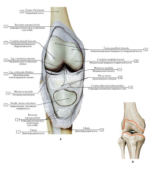 Рис. 222. Коленный сустав, правый (А - вид спереди, суставная капсула вскрыта, Б - места прикрепрепления суставной капсулы на бедренной и большеберцовой костях, вид спереди):
1 - Fibula; 2 - Suprapatellar recess; 3 - Patella, articular surface; 4 - Lateral meniscus; 5 - Fibular collateral ligament; 6 - Anterior cruciate ligament; 7 - Lateral condyle of femur; 8 - Femur; Thigh bone; 9 - Patellar surface of femur; 10 - Medial condyle of femur; 11 - Medial meniscus; 12 - Alar folds; 13 - Infrapatellar fat pad; 14 - Fibula
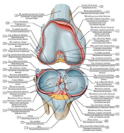 Рис. 223. Коленный сустав, правый, вид спереди и сверху:
1 - Tibia; 2 - Patellar ligament; 3 - Fibula; 4 - Infrapatellar fat pad; 5 - Fibrous layer; Fibrous membrane; Joint capsule; Articular capsule; 6 - Superior articular surface; 7 - Lateral meniscus; 8 - Head of fibula; 9 - Subpopliteal recess; 10 - Popliteus; Tendon; 11 - Fibular collateral ligament; 12 - Posterior meniscofemoral ligament; 13 - Lateral condyle; 14 - Iliotibial tract; 15 - Quadriceps femoris; Tendon; 16 - Femur; Thigh bone; 17 - Suprapatellar bursa; 18 - Synovial membrane; Synovial layer; Joint capsule; Articular capsule; 19 - Tibial collateral ligament; 20 - Infrapatellar synovial fold; 21 - Medial condyle; 22 - Posterior cruciate ligament; 23 - Anterior cruciate ligament; 24 - Semimembranosus; Tendon; 25 - Medial meniscus; 26 - Alar folds
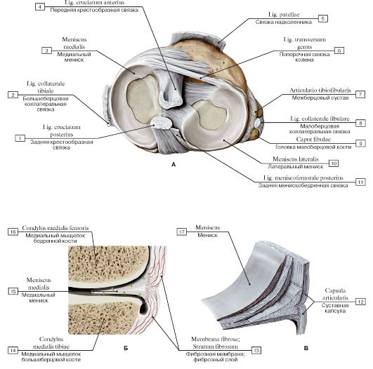 Рис. 224. Мениски коленного сустава, правого (А - вид сверху, Б - медиальный мениск, поперечное сечение, В - мениск, вид изнутри):
1 - Posterior cruciate ligament; 2 - Tibial collateral ligament; 3 - Medial meniscus; 4 - Anterior cruciate ligament; 5 - Patellar ligament; 6 - Transverse ligament of knee; 7 - Tibiofibular joint; Superior tibiofibular joint; 8 - Fibular collateral ligament; 9 - Head of fibula; 10 - Lateral meniscus; 11 - Posterior meniscofemoral ligament; 12 - Joint capsule; Articular capsule; 13 - Fibrous layer; Fibrous membrane; 14 - Medial condyle of tibia; 15 - Meniscus; 16 - Medial condyle of femur; 17 - Meniscus
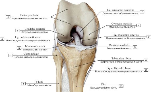 Рис. 225. Крестообразные связки при согнутой голени, правой:
1 - Fibula; 2 - Head of fibula; 3 - Lateral meniscus; 4 - Fibular collateral ligament; 5 - Lateral condyle; 6 - Patellar surface; 7 - Posterior cruciate ligament; 8 - Medial condyle; 9 - Anterior cruciate ligament; 10 - Medial meniscus; 11 - Tibial tuberosity; 12 - Tibial collateral
ligament; 13 - Tibia
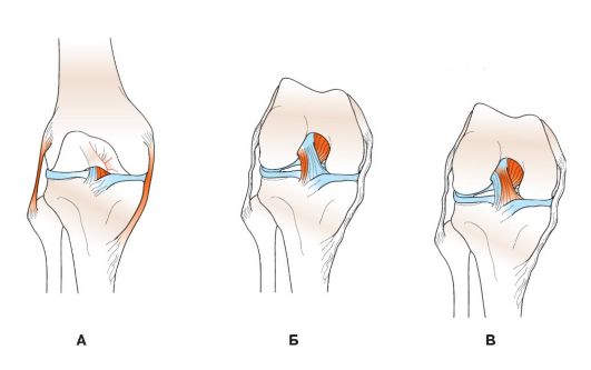 Рис. 226. Роль крестообразных и коллатеральных связок в сгибании и разгибании голени
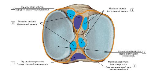 Рис. 227. Места прикрепления менисков, крестообразных связок и синовиальной мембраны на большеберцовой кости,
правой, вид сверху:
1 - Posterior cruciate ligament; 2 - Medial meniscus; 3 - Anterior cruciate ligament; 4 - Lateral meniscus; 5 - Superior articular facet;
6 - Synovial membrane; Synovial layer
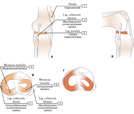 Рис. 228. Движение менисков во время сгибания колена (правый коленный сустав
при разгибании (А) и сгибании (Б), вид сбоку, вид сверху на верхнюю поверхность большеберцовой кости при разгибании (В) и сгибании (Г) голени):
1 - Patella; 2 - Fibular collateral ligament; 3 - Patellar ligament; 4 - Medial meniscus; 5 - Tibial collateral ligament; 6 - Lateral meniscus
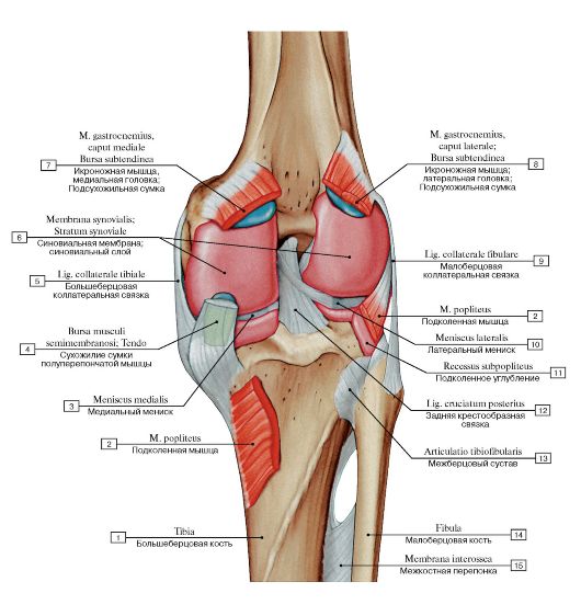 Рис. 229. Синовиальная мембрана коленного сустава, правого, вид сзади:
1 - Tibia; 2 - Popliteus; 3 - Medial meniscus; 4 - Semimembranosus bursa; Tendon; 5 - Tibial collateral ligament; 6 - Synovial membrane; Synovial layer; 7 - Gastrocnemius, medial head; Subtendinous bursa; 8 - Gastrocnemius, lateral head; Subtendinous bursa; 9 - Fibular collateral ligament; 10 - Lateral meniscus; 11 - Subpopliteal recess; 12 - Posterior cruciate ligament; 13 - Tibiofibular joint; Superior tibiofibular joint; 14 - Fibula; 15 - Interosseous membrane
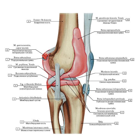 Рис. 230. Связки и сумки коленного сустава, правого, вид с латеральной стороны:
1 - Interosseous membrane of leg; 2 - Fibula; 3 - Tibiofibular joint; Superior tibiofibular joint; 4 - Fibular collateral ligament; 5 - Subpopliteal recess; 6 - Popliteus; Tendon; 7 - Subtendinous bursa; 8 - Gastrocnemius, lateral head; 9 - Femur; Thigh bone; 10 - Quadriceps femoris; Tendon; 11 - Suprapatellar bursa; 12 - Subcutaneous prepatellar bursa; 13 - Lateral meniscus; 14 - Patellar ligament; 15 - Subcutaneous infrapatellar bursa; 16 - Deep infrapatellar bursa; 17 - Synovial membrane; Synovial layer; 18 - Tibia
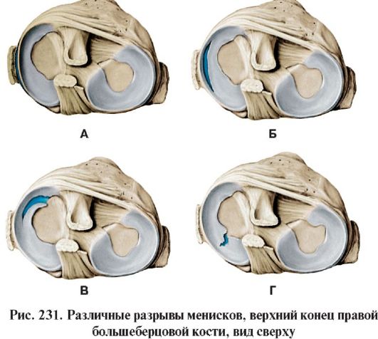 A. Периферический разрыв. стать разрыв мениска или отрыв его от места прикрепле- Б. Разрыв в виде «ручки корзины». ния по периферии. При острой травме мениска возникает
чаще латеральных. Повреждения менисков обычно про- Возрастные дегенеративные изменения в менисках усили-
|
|
Дата добавления: 2014-11-20; Просмотров: 4661; Нарушение авторских прав?; Мы поможем в написании вашей работы!