КАТЕГОРИИ:
Архитектура-(3434)Астрономия-(809)Биология-(7483)Биотехнологии-(1457)Военное дело-(14632)Высокие технологии-(1363)География-(913)Геология-(1438)Государство-(451)Демография-(1065)Дом-(47672)Журналистика и СМИ-(912)Изобретательство-(14524)Иностранные языки-(4268)Информатика-(17799)Искусство-(1338)История-(13644)Компьютеры-(11121)Косметика-(55)Кулинария-(373)Культура-(8427)Лингвистика-(374)Литература-(1642)Маркетинг-(23702)Математика-(16968)Машиностроение-(1700)Медицина-(12668)Менеджмент-(24684)Механика-(15423)Науковедение-(506)Образование-(11852)Охрана труда-(3308)Педагогика-(5571)Полиграфия-(1312)Политика-(7869)Право-(5454)Приборостроение-(1369)Программирование-(2801)Производство-(97182)Промышленность-(8706)Психология-(18388)Религия-(3217)Связь-(10668)Сельское хозяйство-(299)Социология-(6455)Спорт-(42831)Строительство-(4793)Торговля-(5050)Транспорт-(2929)Туризм-(1568)Физика-(3942)Философия-(17015)Финансы-(26596)Химия-(22929)Экология-(12095)Экономика-(9961)Электроника-(8441)Электротехника-(4623)Энергетика-(12629)Юриспруденция-(1492)Ядерная техника-(1748)
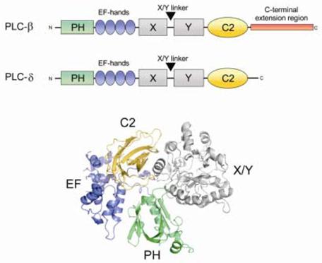
PLC is activated by two G-protein alpha subunits, alpha-q and alpha-11, as well as G-beta gamma subunits
|
|
|
|
This reaction uses calcium as a cofactor and plays an important role in the intracellular transduction of many extracellular signals.

The PLCβ – G protein signaling pathway showing how the binding of an extracellular to a transmembrane GPCR will activate a G protein heterotrimer whose G α or G βγ subunits can bind to and activate PLC β.
Activated PLC β will hydrolyze PI(4,5)P2 releasing Ins(1,4,5)P3 into the cytosol where it diffuses to the endoplasmic reticulum (ER) causing the release of calcium ions into the cytosol. The lipid portion from PIP(4,5)P2 hydrolysis, diacylglyercol, activates protein kinase C (PKC).
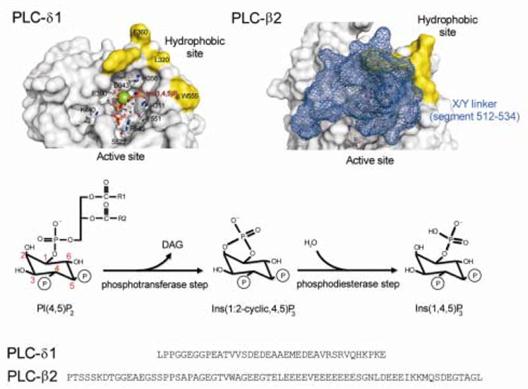
Comparison of the structure of the PH domains of the PLC-δ1 and –β2
Top left – The structure of rat PH-δ1. Top right - The structure of the human PH-β2. Bottom - The structural alignment of PH-δ1 and PH-β2 sequences showing the absence of the Ins(1,4,5)P3 binding residues and the longer β5/β6 loop of PH-β2.
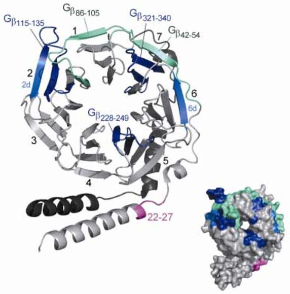
PLC-β2 binding sites on Gβγ
The region corresponding to the signal transfer regions are represented in light green whereas the general PLC-β2 binding domain are in dark blue. Additional regions found to be important for the Gβγ -activation of PLC-β2 are in light blue. The second-binding site for PLC-β2 found in the N-terminal region of Gβ are in magenta.
The Gγ subunit is colored in dark grey. The numeration of blade of the β-propeller is indicated.
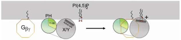
A model of Gβγ activation of PLC-β2
The PH domain has an inhibitory effect on the X/Y domain through the residues 71–88 (β5/β6 loop) by maintaining the catalytic domain in a non- productive orientation.
Gβγ (by binding to the PH and X/Y domain) induces a slight conformational change allowing the X/Y domain to be correctly positioned at the membrane surface and hydrolyze the substrate.
|
|
|
|
|
Дата добавления: 2014-01-14; Просмотров: 573; Нарушение авторских прав?; Мы поможем в написании вашей работы!