КАТЕГОРИИ:
Архитектура-(3434)Астрономия-(809)Биология-(7483)Биотехнологии-(1457)Военное дело-(14632)Высокие технологии-(1363)География-(913)Геология-(1438)Государство-(451)Демография-(1065)Дом-(47672)Журналистика и СМИ-(912)Изобретательство-(14524)Иностранные языки-(4268)Информатика-(17799)Искусство-(1338)История-(13644)Компьютеры-(11121)Косметика-(55)Кулинария-(373)Культура-(8427)Лингвистика-(374)Литература-(1642)Маркетинг-(23702)Математика-(16968)Машиностроение-(1700)Медицина-(12668)Менеджмент-(24684)Механика-(15423)Науковедение-(506)Образование-(11852)Охрана труда-(3308)Педагогика-(5571)Полиграфия-(1312)Политика-(7869)Право-(5454)Приборостроение-(1369)Программирование-(2801)Производство-(97182)Промышленность-(8706)Психология-(18388)Религия-(3217)Связь-(10668)Сельское хозяйство-(299)Социология-(6455)Спорт-(42831)Строительство-(4793)Торговля-(5050)Транспорт-(2929)Туризм-(1568)Физика-(3942)Философия-(17015)Финансы-(26596)Химия-(22929)Экология-(12095)Экономика-(9961)Электроника-(8441)Электротехника-(4623)Энергетика-(12629)Юриспруденция-(1492)Ядерная техника-(1748)
Diagnostics
|
|
|
|
Management Strategy
Threatened Leg
As shown in Table 4, the threatened leg differs from the viable one in that the sensibility is impaired and there is no measurable ankle blood pressure. The threatened limb is further separated into marginally threatened and immediately threatened by the presence or absence of normal motor function. The threatened leg differs from the irreversibly damaged leg by the quality of the venous Doppler signal. In the irreversibly damaged leg, venous blood flow is stagnant and inaudible.
A viable leg does not require immediate action and can be observed in the ward. A threatened leg needs urgent operation or thrombolysis. The latter is more time-consuming and recommended for the marginally threatened leg. The immediately threatened leg must be treated as soon as possible, usually with embolectomy or a vascular reconstruction. Irreversible ischemia is quite unusual but implies that the patient’s leg cannot be saved. Figure 2 is intended to show a simplified algorithm to further support the management of acute leg ischemia.
A well-conducted physical examination is enough to confirm the diagnosis of acute leg ischemia, determine the level of obstruction, and evaluate the severity of ischemia. When the leg is immediately threatened, further radiologic examinations or vascular laboratory tests should not under any circumstances delay surgical treatment. When the extremity is viable or marginally threatened, angiography should be performed. Duplex ultrasound is of limited value for evaluating acute leg ischemia and angiography is recommended for almost all patients in these two groups. If angiography is not available or if examination of the patient has verified that emboli is the cause and probably is best treated by embolectomy, angiography can be omitted. This situation is rare, however.
The arteriogram provides an anatomical map of the vascular bed and is very helpful in discriminating embolus and thrombosis. The former is essential for planning the surgical procedure, and the latter may be of importance for selecting the treatment strategy.
An arteriogram representing an embolus is shown in Fig. 3.
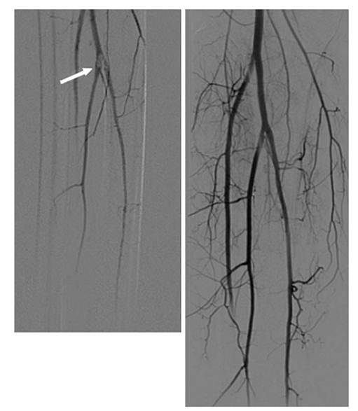
Fig. 3. Embolus lodged at the origins of the calf vessels (arrow).
Angiograms display ilms before and after thrombolysis
Angiographic signs of embolism are an abrupt, convex start of the occlusion and lack of collaterals. Thrombosis is likely when the arteriogram shows well-developed collaterals and atherosclerotic changes in other vascular segments.
For most patients with viable and marginally threatened legs the diagnostic angiography is followed by therapeutic thrombolysis right away.
Angiography can be performed during daytime when qualified radiology staff is available. The patient should be optimized according to the recommendations given in the next section. Before angiography it is important to keep the patient well hydrated and to stop administration of metformin to reduce the risk of renal failure. Disturbances in coagulation parameters may interfere with arterial puncture and must also be checked before the investigation. The information is also important as baseline values in case of later thrombolysis.
The groin of the contralateral leg is the preferred puncture site for diagnostic angiography. A second antegrade puncture can be done in the ischemic extremity if thrombolysis is feasible.
|
|
|
|
|
Дата добавления: 2014-12-23; Просмотров: 358; Нарушение авторских прав?; Мы поможем в написании вашей работы!