КАТЕГОРИИ:
Архитектура-(3434)Астрономия-(809)Биология-(7483)Биотехнологии-(1457)Военное дело-(14632)Высокие технологии-(1363)География-(913)Геология-(1438)Государство-(451)Демография-(1065)Дом-(47672)Журналистика и СМИ-(912)Изобретательство-(14524)Иностранные языки-(4268)Информатика-(17799)Искусство-(1338)История-(13644)Компьютеры-(11121)Косметика-(55)Кулинария-(373)Культура-(8427)Лингвистика-(374)Литература-(1642)Маркетинг-(23702)Математика-(16968)Машиностроение-(1700)Медицина-(12668)Менеджмент-(24684)Механика-(15423)Науковедение-(506)Образование-(11852)Охрана труда-(3308)Педагогика-(5571)Полиграфия-(1312)Политика-(7869)Право-(5454)Приборостроение-(1369)Программирование-(2801)Производство-(97182)Промышленность-(8706)Психология-(18388)Религия-(3217)Связь-(10668)Сельское хозяйство-(299)Социология-(6455)Спорт-(42831)Строительство-(4793)Торговля-(5050)Транспорт-(2929)Туризм-(1568)Физика-(3942)Философия-(17015)Финансы-(26596)Химия-(22929)Экология-(12095)Экономика-(9961)Электроника-(8441)Электротехника-(4623)Энергетика-(12629)Юриспруденция-(1492)Ядерная техника-(1748)
Popliteal Artery Above the Knee, Fig. 6 A, B
|
|
|
|
Superficial Femoral Artery, Fig. 5
A skin incision is made along the dorsal aspect of the sartorius muscle at a midthigh level. It is important to avoid injuries to the greater saphenous vein, which usually is located in the posterior flap of the incision. The incision can be elongated as needed. After the deep fascia is opened and the sartorius muscle is retracted anteriorly, the femoral artery is found and can be mobilized. Division of the adductor tendon is sometimes required for exposure.
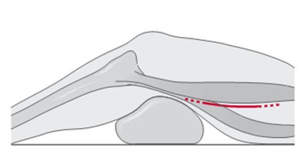
Fig. 5. Incision for exposure of the supericial femoral artery
a The knee is supported on a sterile, draped pillow. The skin incision is started at the medial aspect of the femoral condyle and follows the anterior border of the sartorius muscle 10–15 cm in a proximal direction. Protect the greater saphenous vein and the saphenous nerve during dissection down to the fascia. After dividing the fascia longitudinally, continue the dissection in the groove between the sartorius and gracilis muscles, which leads to the fat in the popliteal fossa.
b The popliteal artery and adjacent veins and nerve are then, without further division of muscles, easily found and separated in the anterior aspect of the fossa.
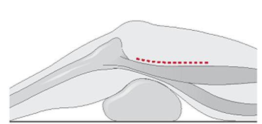
Fig. 6 A. Exposure of popliteal artery above the knee
Fig. 6 B. Exposure of popliteal artery above the knee
|
|
|
|
|
Дата добавления: 2014-12-23; Просмотров: 359; Нарушение авторских прав?; Мы поможем в написании вашей работы!