КАТЕГОРИИ:
Архитектура-(3434)Астрономия-(809)Биология-(7483)Биотехнологии-(1457)Военное дело-(14632)Высокие технологии-(1363)География-(913)Геология-(1438)Государство-(451)Демография-(1065)Дом-(47672)Журналистика и СМИ-(912)Изобретательство-(14524)Иностранные языки-(4268)Информатика-(17799)Искусство-(1338)История-(13644)Компьютеры-(11121)Косметика-(55)Кулинария-(373)Культура-(8427)Лингвистика-(374)Литература-(1642)Маркетинг-(23702)Математика-(16968)Машиностроение-(1700)Медицина-(12668)Менеджмент-(24684)Механика-(15423)Науковедение-(506)Образование-(11852)Охрана труда-(3308)Педагогика-(5571)Полиграфия-(1312)Политика-(7869)Право-(5454)Приборостроение-(1369)Программирование-(2801)Производство-(97182)Промышленность-(8706)Психология-(18388)Религия-(3217)Связь-(10668)Сельское хозяйство-(299)Социология-(6455)Спорт-(42831)Строительство-(4793)Торговля-(5050)Транспорт-(2929)Туризм-(1568)Физика-(3942)Философия-(17015)Финансы-(26596)Химия-(22929)Экология-(12095)Экономика-(9961)Электроника-(8441)Электротехника-(4623)Энергетика-(12629)Юриспруденция-(1492)Ядерная техника-(1748)
Lymphatic Malformation
|
|
|
|
Lymphatic malformations (LMs) are frequently called "lymphangiomas," erroneously suggesting a proliferative tendency. LMs are not tumors. Their pathogenesis is unknown. LMs of the neck and axilla may be due to failure of lymphatic sacs to communicate with the central venous system. Sequestration of lymphatic buds may account for peripheral lesions. Typical LMs seem to be sporadic. Heritable forms of lymphedema, also a type of LM, have been identified.
LMs are classified as microcystic, macrocystic, or combined macrocystic and microcystic lesions.
The difference is clinically and therapeutically useful. Size is distinguished by whether or not the cystic cavity can be successfully aspirated, resulting in visible decompression. LMs may be focal, discrete masses or infiltrate multiple anatomic locations.
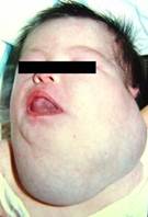
LMs are usually apparent at birth and tend to occur in areas of major lymphatic channels, especially the cervical and axillary locations (Image 9.6). They can be found in all tissues or organ systems with the exception of the central nervous system. LMs most commonly appear as ballottable masses with normal overlying skin, although a blue hue may result if large underlying cysts are present.
LM is most commonly located in the head, neck, axilla, and mediastinum, and may be localized or affect entire organ systems.
Histologically, LMs appear as thin-walled vascular channels lined by lymphatic endothelial cells, which are immunopositive for podoplanin (D2-40) and LYVE-1. The lumens may be empty or filled with a proteinaceous fluid containing macrophages and lymphocytes.
 A
A
|  B
B
|
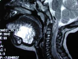
| Image 9.6 A – Cervical lymphatic malformation; B – MRI of LM lesions. |
LM is a slow-flow anomaly that is usually noted at birth or prior to 2 years of age. LM may be noted on prenatal ultrasound during the last trimester. Occasionally, LM presents in late childhood or adulthood after bleeding or infection. Although most LM are sporadic, midline LM can be associated with chromosomal aneuploidy. Posterior cervical LM has been linked to trisomy 13, 18, and 21 and to Roberts and Noonan syndromes. The hereditary form of lymphedema (Milroy syndrome), a type of LM, has been linked to chromosomes 5q and 16q.
|
|
|
|
|
Дата добавления: 2014-10-15; Просмотров: 268; Нарушение авторских прав?; Мы поможем в написании вашей работы!