КАТЕГОРИИ:
Архитектура-(3434)Астрономия-(809)Биология-(7483)Биотехнологии-(1457)Военное дело-(14632)Высокие технологии-(1363)География-(913)Геология-(1438)Государство-(451)Демография-(1065)Дом-(47672)Журналистика и СМИ-(912)Изобретательство-(14524)Иностранные языки-(4268)Информатика-(17799)Искусство-(1338)История-(13644)Компьютеры-(11121)Косметика-(55)Кулинария-(373)Культура-(8427)Лингвистика-(374)Литература-(1642)Маркетинг-(23702)Математика-(16968)Машиностроение-(1700)Медицина-(12668)Менеджмент-(24684)Механика-(15423)Науковедение-(506)Образование-(11852)Охрана труда-(3308)Педагогика-(5571)Полиграфия-(1312)Политика-(7869)Право-(5454)Приборостроение-(1369)Программирование-(2801)Производство-(97182)Промышленность-(8706)Психология-(18388)Религия-(3217)Связь-(10668)Сельское хозяйство-(299)Социология-(6455)Спорт-(42831)Строительство-(4793)Торговля-(5050)Транспорт-(2929)Туризм-(1568)Физика-(3942)Философия-(17015)Финансы-(26596)Химия-(22929)Экология-(12095)Экономика-(9961)Электроника-(8441)Электротехника-(4623)Энергетика-(12629)Юриспруденция-(1492)Ядерная техника-(1748)
Clinical Features
|
|
|
|
Venous Malformation
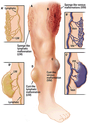
Venous malformations (VMs) are slow-flow lesions that have incorrectly been referred to as “cavernous hemangiomas” in the past. They may be seen at birth or become apparent later depending on location. VMs are most common in the skin and soft tissues but can be located anywhere in the body (Image 9.7).
|
| Image 9.7 Venous malformations and Lymphatic malformations |
Most VMs are sporadic (90%). Somatic mutations in the tyrosine kinase receptor TIE2 have been discovered in approximately 50% of sporadic VMs. An autosomal dominant inheritance pattern occurs in cutaneomucosal venous malformation, which accounts for only 1% to 2% of lesions. Mutations in the TEK gene, which encodes TIE2, have been identified.
Familial multiple glomangioma is an autosomal dominant disorder with multiple dermal lesions usually affecting the lower extremity. Histologically, glomangiomas have glomus cells lining the venous channels, unlike the typical VM. Familial cutaneous-mucosal VM is an autosomal dominant syndrome characterized by several small dome-shaped lesions. VM may also occur in patients with Turner's syndrome.
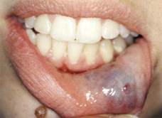
VM is present at birth, but may not become clinically evident until later in life. Although most VMs are located in the skin or subcutaneous tissue, VM also may be located in other organs. They are bluish and compressible, and may be localized, extensive, solitary, or multiple (Image 9.8).
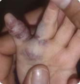
The lesions are mostly superficial and occur as multiple blue to deep-purple nodules or confluent, cobblestone-appearing plaques frequently on the trunk or extremities. They are caused by loss-of-function mutations in glomulin, which derails vascular smooth muscle cell differentiation.
| 
|
| Image 9.8 Venous malformations. |
Diagnosis is facilitated by demonstrating expansion when the affected area is dependent or after a Valsalva maneuver (for head and neck lesions). Phlebothrombosis can cause lesions to become hard and painful. VM expands slowly and usually grows proportionately with the child. Puberty can exacerbate the growth of VM.
|
|
|
|
|
Дата добавления: 2014-10-15; Просмотров: 319; Нарушение авторских прав?; Мы поможем в написании вашей работы!