КАТЕГОРИИ:
Архитектура-(3434)Астрономия-(809)Биология-(7483)Биотехнологии-(1457)Военное дело-(14632)Высокие технологии-(1363)География-(913)Геология-(1438)Государство-(451)Демография-(1065)Дом-(47672)Журналистика и СМИ-(912)Изобретательство-(14524)Иностранные языки-(4268)Информатика-(17799)Искусство-(1338)История-(13644)Компьютеры-(11121)Косметика-(55)Кулинария-(373)Культура-(8427)Лингвистика-(374)Литература-(1642)Маркетинг-(23702)Математика-(16968)Машиностроение-(1700)Медицина-(12668)Менеджмент-(24684)Механика-(15423)Науковедение-(506)Образование-(11852)Охрана труда-(3308)Педагогика-(5571)Полиграфия-(1312)Политика-(7869)Право-(5454)Приборостроение-(1369)Программирование-(2801)Производство-(97182)Промышленность-(8706)Психология-(18388)Религия-(3217)Связь-(10668)Сельское хозяйство-(299)Социология-(6455)Спорт-(42831)Строительство-(4793)Торговля-(5050)Транспорт-(2929)Туризм-(1568)Физика-(3942)Философия-(17015)Финансы-(26596)Химия-(22929)Экология-(12095)Экономика-(9961)Электроника-(8441)Электротехника-(4623)Энергетика-(12629)Юриспруденция-(1492)Ядерная техника-(1748)
Symptoms
|
|
|
|
Arteriovenous malformations
Arteriovenous malformations (AVMs) are fast-flow malformations characterized by abnormal collections of arteries and veins that directly communicate (shunts), thus bypassing the high-resistance capillary bed (Image 9.9). The shunts comprise the epicenter of the AVM, called the nidus. Intracranial AVMs are more common than extracranial AVMs.
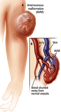
| Image 9.9 Arteriovenous malformations. |
Areas affected by extracranial AVMs in decreasing frequency are the head and neck, limbs, trunk, and viscera. AVMs may be apparent at birth but are often misdiagnosed initially as a capillary malformation or infantile hemangioma due to pink staining in the overlying skin.
Although arteriovenous malformations are present in neonates at birth, they often suddenly become obvious when the patient is older because of various stimuli such as trauma, pregnancy, or puberty. Progression may also occur following biopsy or surgical intervention (e.g., ligation, partial surgical excision).
Common symptoms include pain, overgrowth of the involved body part, changes related to decreased blood flow (ischemia), bleeding, and heart failure. Bleeding is usually minor, but it may be very serious; it typically occurs with dental work in patients with arteriovenous malformation of the dental arcade. Schobinger's staging (stages 1-4) is commonly used to describe the degree of progression (Table 9.2).
Table 9.2
| Clinical Staging System for Arteriovenous Malformation | |
| Stage | Clinical Findings |
| I (Quiescence) | Pink-blue warm stain, shunting on Doppler examination |
| II (Expansion) | Enlargement, pulsation, thrill, bruit, tense veins |
| III (Destruction) | Dystrophic skin changes, ulceration, bleeding, pain, or tissue necrosis |
| IV (Decompensation) | Cardiac failure |
As an AVM grows, it can become more masslike, causing ulceration of the overlying soft tissue, bleeding, pain, or heart failure. Lower-extremity AVMs often develop curious, dry, brown-violaceous-colored plaques.
|
|
|
|
|
Дата добавления: 2014-10-15; Просмотров: 310; Нарушение авторских прав?; Мы поможем в написании вашей работы!