КАТЕГОРИИ:
Архитектура-(3434)Астрономия-(809)Биология-(7483)Биотехнологии-(1457)Военное дело-(14632)Высокие технологии-(1363)География-(913)Геология-(1438)Государство-(451)Демография-(1065)Дом-(47672)Журналистика и СМИ-(912)Изобретательство-(14524)Иностранные языки-(4268)Информатика-(17799)Искусство-(1338)История-(13644)Компьютеры-(11121)Косметика-(55)Кулинария-(373)Культура-(8427)Лингвистика-(374)Литература-(1642)Маркетинг-(23702)Математика-(16968)Машиностроение-(1700)Медицина-(12668)Менеджмент-(24684)Механика-(15423)Науковедение-(506)Образование-(11852)Охрана труда-(3308)Педагогика-(5571)Полиграфия-(1312)Политика-(7869)Право-(5454)Приборостроение-(1369)Программирование-(2801)Производство-(97182)Промышленность-(8706)Психология-(18388)Религия-(3217)Связь-(10668)Сельское хозяйство-(299)Социология-(6455)Спорт-(42831)Строительство-(4793)Торговля-(5050)Транспорт-(2929)Туризм-(1568)Физика-(3942)Философия-(17015)Финансы-(26596)Химия-(22929)Экология-(12095)Экономика-(9961)Электроника-(8441)Электротехника-(4623)Энергетика-(12629)Юриспруденция-(1492)Ядерная техника-(1748)
Acute intestinal ischemia
|
|
|
|
Mesenteric artery thrombosis has the highest mortality rate of all causes of mesenteric ischemia. First described in the late 15th century, little progress was made in its treatment before the 20th century.
In 1901, a patient with a long history of postprandial pain was found to have an atherosclerotic plaque with overlying thrombus of the superior mesenteric artery (SMA). The physician concluded that if a patient could develop pain of the lower extremities secondary to atherosclerosis, it would stand to reason that a patient could present with postprandial pain due to narrowing of the mesenteric vessels. An example of complete occlusion is illustrated in the image below. The pathophysiologic mechanism by which ischemia produces pain remains poorly understood.
The arterial circulation to the gut has extensive collaterals and arcades providing multiple sources of blood inflow. This explains why vascular occlusion is well tolerated as evidenced by the relative lack of clinical intestinal ischemia despite the high prevalence of atherosclerotic disease of the aorta and visceral arteries. Certain collateral patterns are recognized, depending on which artery is blocked. When either the celiac or superior mesenteric artery (SMA) is compromised, the main collateral circulation is by the gastroduodenal and pancreaticoduodenal arteries. The main collateral channels between the SMA and inferior mesenteric artery (IMA) occur in the region of the splenic flexure between the middle and left colic arteries. In the presence of either SMA or IMA occlusion, the marginal artery of Drummond and the arch of Riolan (an ascending branch of the left colic artery anastomosing with branches of the SMA) enlarge significantly. In the presence of an IMA occlusion, another important collateral circulation is between the internal iliac artery and the left colic artery via the superior hemorrhoidal arteries.
The SMA is the critically important vessel in maintaining visceral perfusion, as demonstrated by increased blood flow after eating. This is not seen in the celiac artery. In chronic ischemia, all patients have SMA stenosis or occlusion, in addition to celiac artery and/or IMA involvement.
Etiology
OCCLUSIVE DISEASE
Emboli. The SMA is the most common site of embolic occlusion although the celiac artery can be affected. There is classically an underlying cardiac problem giving rise to the organized thrombus that embolizes. This is usually atrial fibrillation or less commonly a mural thrombus from an acute myocardial infarction. A history of previous embolic events is not uncommon. Other causes of emboli include iatrogenic intra-aortic manipulations, paradoxical emboli through a septal defect, atrial myxoma or primary aortic tumors.
The history is of constant severe epigastric or periumbilical pain of sudden onset. It is frequently followed by copious vomiting and explosive diarrhea.
Typically the patient has been previously well and asymptomatic. The abdominal signs are often lacking or nonspecific, with distension in association with absent or normal bowel sounds without any signs of peritonism. This combination of severe abdominal pain out of proportion to the clinical findings is typical. Peritonism or blood in the stool or vomitus indicates severe advanced intestinal ischemia with likely infarction and is generally a late clinical feature.
|
|
|
The presence of proximal SMA pulsation and the distribution of intestinal ischemia are intra-operative clues for an embolus. The occlusion in embolism is usually distal to the origin of the pancreaticoduodenal and middle colic branches, which allows some blood flow to the small intestine to be maintained. The stomach, duodenum, and proximal jejunum are normal with ischemia extending to the mid transverse colon.
Thrombosis.
Thrombosis of the superior mesenteric or celiac arteries is most often associated with a preexisting atherosclerotic lesion that already compromises flow. The most common preexisting pathology found in patients with acute mesenteric thrombosis is atherosclerosis.
Many patients present with histories consistent with chronic mesenteric ischemia. Wasting, postprandial pain, and phagophobia (fear of eating) are all common.
Typically, the atherosclerotic lesion gradually compromises flow to the gut, causing a progressive worsening of symptoms. During a period of low flow, the artery thromboses, and flow to the gut is compromised.
Unlike embolic events that occur in arterial branches and result in limited bowel ischemia, thrombosis occurs at the vessel origin, resulting in extensive bowel involvement.
Superior mesenteric arterial thrombosis may occur as the result of progression of SMA stenosis that had not previously been diagnosed or treated. There is often a history of intestinal or food fear with severe weight loss, the hallmark of chronic intestinal ischemia in about 65% of patients. The typical patient is female and a heavy smoker, often with evidence of widespread arterial disease including previous myocardial infarction or daudication. As with embolic occlusion, the combination of severe abdominal pain out of proportion to the clinical findings is typical. The thrombosis of the SMA occurs at the origin of the artery.
In contrast to embolic disease, the proximal SMA pulse is absent and the distribution of intestinal ischemia is more extensive. Only the stomach, duodenum and distal colon are spared.
In the young patients, fibromuscular dysplasia can cause mesenteric arterial thrombosis with equally devastating results. Intravenous cocaine abuse is another increasing problem accounting for intestinal ischemia in the young patients. The extent of intestinal ischemia and infarction tends to be foca and less than that seen with atherosclerotic thrombosis. The mechanism of ischemia appears to be occlusive rather than due to vasospasm. Mesenteric ischemia should be considered in the differential diagnosis when evaluating a young patient with a history of cocaine abuse presenting with an acute abdomen.
Some prothrombotic states such as hyperhomocysteinemia or the 20210 A prothrombin gene mutation have resulted in primary arterial thrombosis.
Mesenteric venous thrombosis (MVT) is rare and accounts for 5% to 15% of all acute mesenteric ischemia. It is classified as primary (where no cause is recognized) or secondary. Secondary MVT may follow hypercoagulable states, portal venous stasis and hypertension, intra-abdominal infection and inflammation or malignancy, use of oral contraceptives and splenectomy. Long-term anticoagulation is required for MVT, because of the high recurrence rates. The clinical presentation is usually less acute than that of arterial occlusion.
|
|
|
Severe but vague abdominal pain that tends to be colicky and slowly progressive is usually present. Few abdominal signs are present except tenderness, distension and decreased bowel sounds. The pain is out of proportion to the physical findings. Fecal occult blood is present in the majority of patients.
There is a pyrexia of greater than 38 °C in 25% to 50% of patients, and 20% have a tachycardia. Leucocytosis ranges from 12000 to 29000.
Frank peritonitis is seen only when transmural infarction or perforation has occurred.
Surgical findings include blood-stained free peritoneal fluid at laparotomy. The affected bowel is cyanotic and edematous with a rubbery texture.
Mesenteric arterial pulsations are present but the veins contain fresh thrombus that extrude when the veins are cut. Infarction is most common in the mid small bowel.


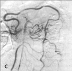
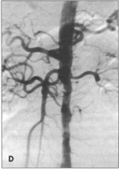
FIG. A - Schematic representation of the collateral circulation of the
intestine. B - Angiosraphic appearance of arch of Riolan from superior
mesenteric artery (stented at its origin). C - Angiographic appearance of
marginal artery of Drummond. D - Initial angiogram demonstrates occlu-
ded IMA. The delayed film shows the colonic supply.
Diagnosis
Acute intestinal ischemia is a life-threatening surgical emergency, yet can be a difficult diagnosis to make, with delay contributing directly to infarction.
The majority of cases are diagnosed more than 12 hours after the onset of symptoms. Delayed diagnosis accounts for the majority of malpractice claims involving acute mesenteric ischemia in the United States. Diagnosis depends on a high index of suspicion. The main presenting feature is the combination of severe abdominal pain out of proportion to the clinical findings, as discussed above.
Serum levels of lactate and leucocytes are elevated in the majority (65% to 90%) of patients to greater than 50 U/L and 15000/mL, respectively.
Hyperamylasemia is seen in just under half the patients with acute mesenteric ischemia. Elevation of serum inorganic phosphate levels have been proposed as a marker of mesenteric ischemia, as it is extensively found in gut, but this only occurs in 15% to 33% of such patients.
However, in those patients who did have elevated phosphate levels, it predicted extensive injury and poor prognosis. The fibrinolytic marker D-dimer is elevated in thrombo-embolic occlusion of the SMA, although levels are also raised in other conditions of acute bowel ischemia such as strangulation or ruptured aortic aneurysm.
Animal studies have suggested intestinal fatty acid binding protein (I-FABP) as a serum marker reflecting bowel ischemia. Early human studies show promise, as patients with ischemic bowel disease demonstrate significantly higher I-FABP levels than either healthy subjects or patients with acute abdominal pain. Patients with mesenteric infarction had the highest serum I-FABP levels.
Plain radiographs of the abdomen may reveal nonspecific bowel dilatation or, in MVT, wall edema (thumbprinting); or gas in the bowel wall or portal vein. Unfortunately they are not helpful in most cases.
Mesenteric angiography will confirm the diagnosis of arterial occlusion but at the cost of delay in treatment. If there are clear abdominal signs of peritonitism, urgent laparotomy without angiography is the best course of action. In the remainder of patients suspected of acute intestinal ischemia with-out abdominal signs, angiography is indicated with lateral views of the visceral aorta and its branches.
In acute SMA thrombosis, there is usually no visualization of the entire artery because of the ostial nature of the disease, although delayed views may show slow filling of the distal SMA. SMA embolization usually allows visualization of the proximal artery to just beyond the level of the middle colic artery.
|
|
|
|
|
Дата добавления: 2014-12-23; Просмотров: 547; Нарушение авторских прав?; Мы поможем в написании вашей работы!