КАТЕГОРИИ:
Архитектура-(3434)Астрономия-(809)Биология-(7483)Биотехнологии-(1457)Военное дело-(14632)Высокие технологии-(1363)География-(913)Геология-(1438)Государство-(451)Демография-(1065)Дом-(47672)Журналистика и СМИ-(912)Изобретательство-(14524)Иностранные языки-(4268)Информатика-(17799)Искусство-(1338)История-(13644)Компьютеры-(11121)Косметика-(55)Кулинария-(373)Культура-(8427)Лингвистика-(374)Литература-(1642)Маркетинг-(23702)Математика-(16968)Машиностроение-(1700)Медицина-(12668)Менеджмент-(24684)Механика-(15423)Науковедение-(506)Образование-(11852)Охрана труда-(3308)Педагогика-(5571)Полиграфия-(1312)Политика-(7869)Право-(5454)Приборостроение-(1369)Программирование-(2801)Производство-(97182)Промышленность-(8706)Психология-(18388)Религия-(3217)Связь-(10668)Сельское хозяйство-(299)Социология-(6455)Спорт-(42831)Строительство-(4793)Торговля-(5050)Транспорт-(2929)Туризм-(1568)Физика-(3942)Философия-(17015)Финансы-(26596)Химия-(22929)Экология-(12095)Экономика-(9961)Электроника-(8441)Электротехника-(4623)Энергетика-(12629)Юриспруденция-(1492)Ядерная техника-(1748)
Infantile Hemangioma
|
|
|
|
Vascular Tumors
Characteristics of Common Vascular Anomalies
| Vascular Tumor / Infantile Hemangioma | Vascular Malformations |
| · Proliferative · Female to male ratio 3:1 · 30% visible at birth · 70% become apparent during first few weeks of life · Rapid postnatal growth followed by slow involution · Endothelial cell proliferation · Increased mast cells · No coagulation abnormalities · High percentage respond dramatically to corticosteroid treatment in 2 to 3 weeks · Immunopositive for biologic markers (including GLUT1) | · Congenital abnormality with proportional growth · No gender predilection · May expand secondary to sepsis, trauma, or hormonal changes · Normal endothelial cell turnover · Normal mast cell count · Do not involute · Localized consumptive coagulopathy possible · Low-flow: phleboliths, ectatic channels · High-flow: enlarged, tortuous vessels with arteriovenous shunting · No response to corticosteroids or antiangiogenic agents · Immunonegative for hemangioma biologic markers |
The International Society for the Study of Vascular Anomalies (ISSVA) classification of vascular anomalies:
| Vascular tumors · Infantile hemangiomas · Congenital hemangiomas (RICH and NICH) · Tufted angioma (with or without Kasabach-Merritt syndrome) · Kaposiform hemangioendothelioma (with or without Kasabach-Merritt syndrome) · Spindle cell hemangioendothelioma · Other, rare hemangioendotheliomas (epithelioid, composite, retiform, polymorphous, Dabska tumor, lymphangioendotheliomatosis, etc.) · Dermatologic acquired vascular tumors (pyogenic granuloma, targetoid hemangioma, glomeruloid hemangioma, microvenular hemangioma, etc.) |
| Vascular malformations Slow-flow vascular malformations: · Capillary malformation (CM) * Port-wine stain * Telangiectasia * Angiokeratoma · Venous malformation (VM) - Common sporadic VM - Bean syndrome - Familial cutaneous and mucosal venous malformation (VMCM) - Glomuvenous malformation (GVM) (glomangioma) - Maffucci syndrome · Lymphatic malformation (LM) Fast-flow vascular malformations: o Arterial malformation (AM) o Arteriovenous fistula (AVF) o Arteriovenous malformation (AVM) Complex-combined vascular malformations: CVM, CLM, LVM, CLVM, AVM-LM, CM-AVM |
| C - capillary; V - venous; L - lymphatic; AV - arteriovenous; M - malformation. RICH - rapidly involuting congenital hemangioma; NICH - noninvoluting congenital hemangioma. |
Hemangioma is a benign, and usually a self-involuting tumor of the endothelial cells (endothelium) that line blood vessels and is characterised by increased number of normal or abnormal vessels filled with blood.
Hemangioma is the most common neoplasm of infancy. Capillary or strawberry hemangioma has previously been used to describe hemangioma involving the dermis, which appeared red. Hemangiomas deep to the dermis may appear bluish and have been referred to as cavernous hemangioma. The terms capillary and cavernous also have been used to describe CM and VM, respectively. The terms capillary, strawberry, and cavernous confuse diagnosis and should not be used.
Infantile hemangioma (IH) occurs in approximately 4% of white-skinned infants. The incidence is lower in dark-skinned babies. There is a female-to-male preponderance of 3:1. Extremely-low-birth-weight infants (<1000 g) have the highest incidence of IHs, approaching 23%. Additional risk factors include advanced maternal age, multiple gestations, and placental abnormalities.
IHs most often occur as a single cutaneous lesion (80%) with a predilection for the head and neck (60%), trunk (25%), and extremities (20%) (Image 9.2). Multiple tumors are present in up to 20% of patients and, when present, may signal involvement of extracutaneous organs such as the liver or gastrointestinal (GI) tract. Median age of onset is 1 to 2 weeks. A premonitory cutaneous mark such as a pale spot or faint macular stain is present at birth in 30% to 50% of cases. The majority (90%) of IHs are small, localized lesions that do not involve aesthetically or functionally vital structures. Endangering or life-threatening IHs are rare.
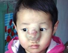
| Image 9.2 Infantile hemangioma. Cervicofacial and subglottic IHs can be life-threatening due to airway obstruction. |
Hemangiomas have unique growth stages:
v a proliferating phase until 1 year of age,
v an involuting phase from 1 to 7 years of age,
v and finally an involuted phase after 7 years of age.
By 5 years of age, 50% of tumors have completed involution, which increases to 70% at 7 years of age. There is often continued gradual regression of the color and bulk of the tumor until 10 to 12 years of age. At the end of involution, 50% of patients have nearly normal skin in the area of the prior lesion. Large tumors can leave lax, redundant skin and/or a fibrofatty residuum. Previously ulcerated lesions can leave permanently damaged skin, scars, and discoloration.
|
|
|
|
|
Дата добавления: 2014-10-15; Просмотров: 503; Нарушение авторских прав?; Мы поможем в написании вашей работы!