КАТЕГОРИИ:
Архитектура-(3434)Астрономия-(809)Биология-(7483)Биотехнологии-(1457)Военное дело-(14632)Высокие технологии-(1363)География-(913)Геология-(1438)Государство-(451)Демография-(1065)Дом-(47672)Журналистика и СМИ-(912)Изобретательство-(14524)Иностранные языки-(4268)Информатика-(17799)Искусство-(1338)История-(13644)Компьютеры-(11121)Косметика-(55)Кулинария-(373)Культура-(8427)Лингвистика-(374)Литература-(1642)Маркетинг-(23702)Математика-(16968)Машиностроение-(1700)Медицина-(12668)Менеджмент-(24684)Механика-(15423)Науковедение-(506)Образование-(11852)Охрана труда-(3308)Педагогика-(5571)Полиграфия-(1312)Политика-(7869)Право-(5454)Приборостроение-(1369)Программирование-(2801)Производство-(97182)Промышленность-(8706)Психология-(18388)Религия-(3217)Связь-(10668)Сельское хозяйство-(299)Социология-(6455)Спорт-(42831)Строительство-(4793)Торговля-(5050)Транспорт-(2929)Туризм-(1568)Физика-(3942)Философия-(17015)Финансы-(26596)Химия-(22929)Экология-(12095)Экономика-(9961)Электроника-(8441)Электротехника-(4623)Энергетика-(12629)Юриспруденция-(1492)Ядерная техника-(1748)
Therapy
|
|
|
|
Diagnosis
The high-flow nature of the malformation can be easily confirmed with Doppler US examination, which reveals high-flow low-resistance arteries and an arterialized waveform in the draining veins.
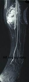
On MRIs, the anomaly is characterized by enlarged vascular channels associated with dilated feeding and draining vessels. A discrete soft-tissue mass is typically absent.
|
| Image 9.10 MR Angiography |
Today, most AVMs are studied with MR Angiography (Image 9.10) before embolization or surgery.
Angiographic embolization alone or in combination with surgical excision is the mainstay of treatment.
Preoperative embolization decreases intraoperative blood loss but does not decrease the extent of resection. Complete removal of the nidus and overlying soft tissue and skin is the goal; this will decrease recurrence.
Controversy exists about when to intervene. In the early stages, the full extent of the lesion may not be appreciated at the time of resection, which could result in local recurrence and complicate future procedures. Meanwhile, a well-localized stage I AVM is often more amenable to resection and complete removal. During infancy, treatment is rarely indicated except in rare cases of postnatal heart failure. After a complete diagnostic evaluation, infants and children should be followed annually.
Treatment is usually delayed until symptoms indicative of stage III develop: tissue destruction, pain, bleeding, or ulceration.
|
|
|
|
|
Дата добавления: 2014-10-15; Просмотров: 309; Нарушение авторских прав?; Мы поможем в написании вашей работы!