КАТЕГОРИИ:
Архитектура-(3434)Астрономия-(809)Биология-(7483)Биотехнологии-(1457)Военное дело-(14632)Высокие технологии-(1363)География-(913)Геология-(1438)Государство-(451)Демография-(1065)Дом-(47672)Журналистика и СМИ-(912)Изобретательство-(14524)Иностранные языки-(4268)Информатика-(17799)Искусство-(1338)История-(13644)Компьютеры-(11121)Косметика-(55)Кулинария-(373)Культура-(8427)Лингвистика-(374)Литература-(1642)Маркетинг-(23702)Математика-(16968)Машиностроение-(1700)Медицина-(12668)Менеджмент-(24684)Механика-(15423)Науковедение-(506)Образование-(11852)Охрана труда-(3308)Педагогика-(5571)Полиграфия-(1312)Политика-(7869)Право-(5454)Приборостроение-(1369)Программирование-(2801)Производство-(97182)Промышленность-(8706)Психология-(18388)Религия-(3217)Связь-(10668)Сельское хозяйство-(299)Социология-(6455)Спорт-(42831)Строительство-(4793)Торговля-(5050)Транспорт-(2929)Туризм-(1568)Физика-(3942)Философия-(17015)Финансы-(26596)Химия-(22929)Экология-(12095)Экономика-(9961)Электроника-(8441)Электротехника-(4623)Энергетика-(12629)Юриспруденция-(1492)Ядерная техника-(1748)
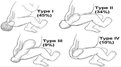
Differential diagnosis. SCT are classified based on the location of the lesion (Image 9.20)
|
|
|
|
Diagnosis
Classification
SCT are classified based on the location of the lesion (Image 9.20). Type I lesions are external with a small presacral component (45% of reported cases). Type II lesions have an external and a significant presacral component (34%). Type III lesions have a small external component with the majority of the tumor being retroperitoneal (9%). Type IV lesions have no external component, being entirely presacral (10%).
|
| Image 9.20 Classification of sacrococcygeal teratomas based on Altman's study. |
The diagnosis of SCT is usually made by physical examination. In neonates presenting with large external masses, the degree of pelvic and abdominal involvement should be assessed preoperatively with ultrasonography, CT, or magnetic resonance imaging (MRI), and these studies may also offer a clue as to the characteristics of the vascular supply.
A chest x-ray is obtained to rule out metastatic disease. Ultrasonography is useful to determine the nature of the lesion (solid vs. cystic), and the presence of liver involvement.
Alpha-fetoprotein (AFP) and beta human chorionic gonadotropin (beta-hCG) are serum tumor markers associated with teratomas and should be obtained preoperatively and followed postoperatively.
The differential diagnosis is quite long and includes lipoma, myelocystocele, pilonidal cysts, sacral dimple, diastematomyelia, meningocele, epidermal sinus, sacral agenesis, fetus in fetu, parasitic twin, hamartoma, hemangioma, neuroblastoma, chordoma, rectal duplication, and sarcoma.
|
|
|
|
|
Дата добавления: 2014-10-15; Просмотров: 292; Нарушение авторских прав?; Мы поможем в написании вашей работы!