КАТЕГОРИИ:
Архитектура-(3434)Астрономия-(809)Биология-(7483)Биотехнологии-(1457)Военное дело-(14632)Высокие технологии-(1363)География-(913)Геология-(1438)Государство-(451)Демография-(1065)Дом-(47672)Журналистика и СМИ-(912)Изобретательство-(14524)Иностранные языки-(4268)Информатика-(17799)Искусство-(1338)История-(13644)Компьютеры-(11121)Косметика-(55)Кулинария-(373)Культура-(8427)Лингвистика-(374)Литература-(1642)Маркетинг-(23702)Математика-(16968)Машиностроение-(1700)Медицина-(12668)Менеджмент-(24684)Механика-(15423)Науковедение-(506)Образование-(11852)Охрана труда-(3308)Педагогика-(5571)Полиграфия-(1312)Политика-(7869)Право-(5454)Приборостроение-(1369)Программирование-(2801)Производство-(97182)Промышленность-(8706)Психология-(18388)Религия-(3217)Связь-(10668)Сельское хозяйство-(299)Социология-(6455)Спорт-(42831)Строительство-(4793)Торговля-(5050)Транспорт-(2929)Туризм-(1568)Физика-(3942)Философия-(17015)Финансы-(26596)Химия-(22929)Экология-(12095)Экономика-(9961)Электроника-(8441)Электротехника-(4623)Энергетика-(12629)Юриспруденция-(1492)Ядерная техника-(1748)
Femoral Artery in the Groin, Fig. 4 A, B, C
|
|
|
|
Exposure of Different Vessel Segments in the Leg
Operation
Threatened Leg
If the leg is immediately threatened, the patient is prepared for operation right away. This includes the steps listed above for the viable leg, including contact with an anesthesiologist. When there is no cyanosis and motor function is normal – that is, the extremity is only marginally threatened – there is time for immediate angiography followed by thrombolysis or operation. An option is cautious monitoring and angiography as soon as possible.
Before starting the operation, the surgeon needs to consider the risk for having to perform a complete vascular reconstruction. It is probable that a bypass to the popliteal artery or a calf artery will be needed to restore circulation. If thrombosis is the likely cause and the obstruction is distal (a palpable pulse is felt in the groin but not distally), a bypass may also be required even when embolization is suspected.
a A longitudinal skin incision starting 1–2 cm cranial to the inguinal skin fold and continued lateral to the artery is used to avoid the inguinal lymph nodes. A common mistake is to place the incision too far caudally, which usually means the dissection is taking place distal to the deep femoral.
b The dissection is continued sharply with the knife straight down to the fascia lateral to the lymph nodes and is then angulated 90° medially to reach the area over the artery. It should then be palpable. Lymph nodes should be avoided to minimize the risk for infection and development of seroma. The fascia is incised, and the anterior and lateral surfaces of the artery are approached.
c At this stage the anatomy is often unclear regarding the relation of branches to the common femoralartery. Encircle the exposed artery with a vessel-loop, and gently lift the artery. Continue dissection until the bifurcation into superficial and deep femoral artery is identified. Its location varies from high up under the inguinal ligament up to 10 cm further down. At this stage, the surgeon must decide whether exposure and clamping of the common femoral are enough. This is usually the case for proximal control in trauma distally in the leg. In acute ischemia it is more common that the entire bifurcation needs to be exposed.
During the continued dissection, attention must be given to important branches that should be controlled and protected from iatrogenic injuries. These are, in particular, the circumflex iliac artery on the dorsal aspect of the common femoral artery and the deep femoral vein crossing over the anterior aspect of the deep femoral artery just after its bifurcation. To provide a safe and good exposure of the deep femoral to a level below its first bifurcation, this vein must be divided and suture-ligated. Partial division of the inguinal ligament is occasionally needed for satisfactory exposure.
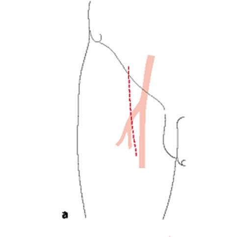
Fig. 4 A. Exposure of femoral artery in the groin
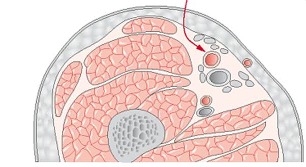
Fig. 4 B. Exposure of femoral artery in the groin
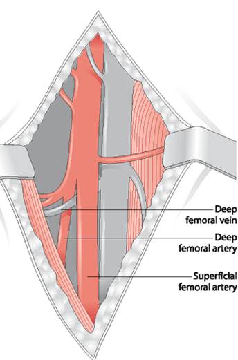
Fig. 4 C. Exposure of femoral artery in the groin
|
|
|
|
|
Дата добавления: 2014-12-23; Просмотров: 410; Нарушение авторских прав?; Мы поможем в написании вашей работы!