КАТЕГОРИИ:
Архитектура-(3434)Астрономия-(809)Биология-(7483)Биотехнологии-(1457)Военное дело-(14632)Высокие технологии-(1363)География-(913)Геология-(1438)Государство-(451)Демография-(1065)Дом-(47672)Журналистика и СМИ-(912)Изобретательство-(14524)Иностранные языки-(4268)Информатика-(17799)Искусство-(1338)История-(13644)Компьютеры-(11121)Косметика-(55)Кулинария-(373)Культура-(8427)Лингвистика-(374)Литература-(1642)Маркетинг-(23702)Математика-(16968)Машиностроение-(1700)Медицина-(12668)Менеджмент-(24684)Механика-(15423)Науковедение-(506)Образование-(11852)Охрана труда-(3308)Педагогика-(5571)Полиграфия-(1312)Политика-(7869)Право-(5454)Приборостроение-(1369)Программирование-(2801)Производство-(97182)Промышленность-(8706)Психология-(18388)Религия-(3217)Связь-(10668)Сельское хозяйство-(299)Социология-(6455)Спорт-(42831)Строительство-(4793)Торговля-(5050)Транспорт-(2929)Туризм-(1568)Физика-(3942)Философия-(17015)Финансы-(26596)Химия-(22929)Экология-(12095)Экономика-(9961)Электроника-(8441)Электротехника-(4623)Энергетика-(12629)Юриспруденция-(1492)Ядерная техника-(1748)
Intraoperative angiography
|
|
|
|
Thrombosis
The preliminary diagnosis of embolus must be reconsidered if the exposed femoral artery in the groin is hard and calcified. In most situations, clot removal with Fogarty catheters will then fail. It is usually difficult or even impossible to pass the catheter distally, indicating the presence of stenoses or occlusions. Even if the embolectomy appears successful, early reocclusion is common. Such secondary thrombosis is usually more extensive and will aggravate the ischemia. Accordingly, angiography should be considered as the first step if the femoral artery is grossly arteriosclerotic and if it is hard to pass the catheter down to the calf level. It will confirm the etiology and reveal whether a bypass is required and feasible. Vascular reconstruction in acute leg ischemia is often rather difficult andexperience in vascular surgery is required.
With the proximal clamp in position a 5 or 8French baby-feeding catheter is inserted into the arteriotomy. The tip of the catheter is placed 5cm into the superficial femoral artery and distal control around it is achieved with a vessel-loop. Contrast for intravasal use containing 140–300 mg iodine/ml is infused with a 20ccsyringe connected to a three-way valve. Heparinized Ringer’s or saline (10units/ml heparin) is flushed through the catheter before and after contrast injection to prevent thrombosis in the occluded vascular bed. If the patient is suspected to have renal failure, the amount of contrast used is kept at a minimum. Angled projections can be obtained without moving the C-arm by rotating the patient’s foot.
The use of contrast in the Fogarty catheter balloon during fluoroscopy allows the calf vessel into which the catheter slides to be identified. The technique for intraoperative angiography is also a prerequisite for interoperative use of endovascular treatment options such asangioplasty (Fig. 9).
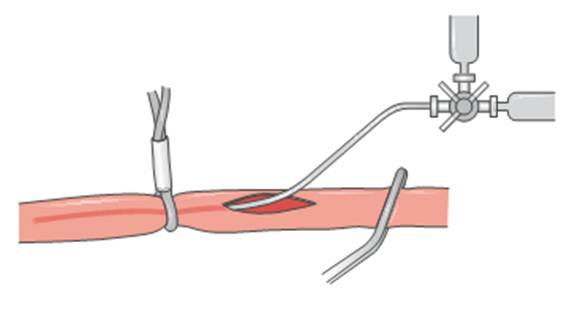
Fig. 9. Intraoperative angiography
|
|
|
|
|
Дата добавления: 2014-12-23; Просмотров: 465; Нарушение авторских прав?; Мы поможем в написании вашей работы!